

DPRC Hospital & Diagnostic Lab
- 29 Probal Housing, Ring Road, Mohammadpur. (Opposite to Suchona Community Center), Dhaka, Dhaka-1207, Bangladesh
- 01716306913, 017132200
- dprclab786@gmail.com
About Us & What we do
Details
DPRC is the largest with different site branches, well-known, registered best privet physiotherapy centers in Bangladesh. The DPRC works with governments and educational bodies to try to ensure that a quality physiotherapy service is available when and where you need it. We know just how important it is for people to keep moving right throughout their life. Our goal is to provide a healing environment and practice physiotherapy at the highest possible level.People often suffer from back, neck, knee & shoulder pain silently. No matter what you are experiencing, we have the right Physiotherapy treatment that can help you. If you are suffering from back, neck, knee & shoulder pain and want to get rid of it, you need to try May physiotherapy treatment. Your health is very important to us and that’s why we always do our best to help many patients.
Our physiotherapy treatment is far advanced than others. If you have back, neck, & shoulder pain questions, we would be glad to answer for you. Whether you need help or information we can help you for any pain problem. Find out more about your pain paralysis and rehabilitation with our online system.
The good news is that most back problems can be treated with no surgical method.
Our Services
Arthritis (from Greek arthro-, joint + -itis, inflammation; plural: arthritides) is a form of joint disorder that involves inflammation of one or more joints. There are over 100 different forms of arthritis. The most common form, osteoarthritis (degenerative joint disease), is a result of trauma to the joint, infection of the joint, or age. Other arthritis forms are rheumatoid arthritis, psoriatic arthritis, and related autoimmune diseases. Septic arthritis is caused by joint infection.
The major complaint by individuals who have arthritis is joint pain. Pain is often a constant and may be localized to the joint affected. The pain from arthritis is due to inflammation that occurs around the joint, damage to the joint from disease, daily wear and tear of joint, muscle strains caused by forceful movements against stiff painful joints and fatigue.
The major complaint by individuals who have arthritis is joint pain. Pain is often a constant and may be localized to the joint affected. The pain from arthritis is due to inflammation that occurs around the joint, damage to the joint from disease, daily wear and tear of joint, muscle strains caused by forceful movements against stiff painful joints and fatigue.
Back pain is pain felt in the back that usually originates from the muscles, nerves, bones, joints or other structures in the spine.
Back pain may have a sudden onset or can be a chronic pain; it can be constant or intermittent, stay in one place or radiate to other areas. It may be a dull ache, or a sharp or piercing or burning sensation. The pain may radiate into the arms and hands as well as the legs or feet, and may include symptoms other than pain. These symptoms may include tingling, weakness or numbness.
Back pain is one of humanity’s most frequent complaints. Acute low back pain (also called lumbago) is the fifth most common reason for physician visits. About nine out of ten adults experience back pain at some point in their life, and five out of ten working adults have back pain every year. Back pain is second only to upper respiratory conditions as the stated cause of work loss. Also, it’s the single leading cause of disability worldwide as Americans spend at least $50 billion each year treating it.
The spine is a complex interconnecting network of nerves, joints, muscles, tendons and ligaments, all of which are capable of producing pain. Large nerves that originate in the spine and go to the legs and arms can make pain radiate to the extremities.
Back pain may have a sudden onset or can be a chronic pain; it can be constant or intermittent, stay in one place or radiate to other areas. It may be a dull ache, or a sharp or piercing or burning sensation. The pain may radiate into the arms and hands as well as the legs or feet, and may include symptoms other than pain. These symptoms may include tingling, weakness or numbness.
Back pain is one of humanity’s most frequent complaints. Acute low back pain (also called lumbago) is the fifth most common reason for physician visits. About nine out of ten adults experience back pain at some point in their life, and five out of ten working adults have back pain every year. Back pain is second only to upper respiratory conditions as the stated cause of work loss. Also, it’s the single leading cause of disability worldwide as Americans spend at least $50 billion each year treating it.
The spine is a complex interconnecting network of nerves, joints, muscles, tendons and ligaments, all of which are capable of producing pain. Large nerves that originate in the spine and go to the legs and arms can make pain radiate to the extremities.
Frozen shoulder, medically referred to as adhesive capsulitis, is a disorder in which the shoulder capsule, the connective tissue surrounding the glenohumeral joint of the shoulder, becomes inflamed and stiff, greatly restricting motion and causing chronic pain.
Adhesive capsulitis is a painful and disabling condition that often causes great frustration for patients and caregivers due to slow recovery. Movement of the shoulder is severely restricted. Pain is usually constant, worse at night, and when the weather is colder; and along with the restricted movement can make even small tasks impossible. Certain movements or bumps can cause sudden onset of tremendous pain and cramping that can last several minutes.
This condition, for which an exact cause is unknown, can last from five months to three years or more and is thought in some cases to be caused by injury or trauma to the area. It is believed that it may have an autoimmune component, with the body attacking healthy tissue in the capsule. There is also a lack of fluid in the joint, further restricting movement.
In addition to difficulty with everyday tasks, people who suffer from adhesive capsulitis usually experience problems sleeping for extended periods due to pain that is worse at night and restricted movement/positions. The condition also can lead to depression, pain, and problems in the neck and back.
Risk factors for frozen shoulder include diabetes, stroke, accidents, lung disease, connective tissue disorders, thyroid disease, and heart disease. The condition very rarely appears in people under 40.
Treatment may be painful and taxing and consists of physical therapy, occupational therapy, Chiropractic, medication, massage therapy, hydrodilatation or surgery. A doctor may also perform manipulation under, which breaks up the adhesions and scar tissue in the joint to help restore some range of motion. Pain and inflammation can be controlled with analgesics and NSAIDs. The condition tends to be self-limiting: it usually resolves over time without surgery, but this may take up to two years. Most people regain about 90% of shoulder motion over time. People who suffer from adhesive capsulitis may have extreme difficulty working and going about normal life activities for several months or longer.
Adhesive capsulitis is a painful and disabling condition that often causes great frustration for patients and caregivers due to slow recovery. Movement of the shoulder is severely restricted. Pain is usually constant, worse at night, and when the weather is colder; and along with the restricted movement can make even small tasks impossible. Certain movements or bumps can cause sudden onset of tremendous pain and cramping that can last several minutes.
This condition, for which an exact cause is unknown, can last from five months to three years or more and is thought in some cases to be caused by injury or trauma to the area. It is believed that it may have an autoimmune component, with the body attacking healthy tissue in the capsule. There is also a lack of fluid in the joint, further restricting movement.
In addition to difficulty with everyday tasks, people who suffer from adhesive capsulitis usually experience problems sleeping for extended periods due to pain that is worse at night and restricted movement/positions. The condition also can lead to depression, pain, and problems in the neck and back.
Risk factors for frozen shoulder include diabetes, stroke, accidents, lung disease, connective tissue disorders, thyroid disease, and heart disease. The condition very rarely appears in people under 40.
Treatment may be painful and taxing and consists of physical therapy, occupational therapy, Chiropractic, medication, massage therapy, hydrodilatation or surgery. A doctor may also perform manipulation under, which breaks up the adhesions and scar tissue in the joint to help restore some range of motion. Pain and inflammation can be controlled with analgesics and NSAIDs. The condition tends to be self-limiting: it usually resolves over time without surgery, but this may take up to two years. Most people regain about 90% of shoulder motion over time. People who suffer from adhesive capsulitis may have extreme difficulty working and going about normal life activities for several months or longer.
Elbow pain
The elbow is the joint where three long bones meet in the middle portion of the arm. The bone of the upper arm (humerus) meets the inner bone of the forearm (ulna) and the outer bone of the forearm (radius) to form a hinge joint. The radius and ulna also meet in the elbow to allow for rotation of the forearm. The elbow functions to move the arm like a hinge (forward and backward) and in rotation (twisting outward and inward). The biceps muscle is the major muscle that flexes the elbow hinge. The triceps muscle is the major muscle that extends the elbow hinge. The outer bone of the elbow is referred to as the lateral epicondyle and is a part of the humerus bone. Tendons are attached to this area which can be injured, causing inflammation or tendinitis (lateral epicondylitis, or “tennis elbow”).The inner portion of the elbow is a bony prominence called the medial epicondyle. Additional tendons from the muscles attach here and can be injured, causing medial epicondylitis, “golfer’s elbow.” A fluid-filled sac (bursa), which serves to reduce friction, overlies the tip of the elbow (olecranon bursa). The elbow can be affected by inflammation of the tendons or the bursae (plural for bursa) or conditions that affect the bones and joints, such as fractures, arthritis, or nerve irritation. Joint pain in the elbow can result from injury or disease involving any of these structures.
Knee pain
The knee joint consists of an articulation between four bones: the femur, tibia, fibula and patella. There are four compartments to the knee. These are the medial and lateral tibiofemoral compartments, the patellofemoral compartment and the superior tibiofibular joint. The components of each of these compartments can suffer from repetitive strain, injury or disease. Running long distance can cause pain tothe knee joint as it is high impact exercise.
Injuries
Some common injuries include :-
Sprain (Ligament sprain)
Medial collateral ligament
Lateral collateral ligament
Anterior cruciate ligament
Posterior cruciate ligament
Tear of meniscus
Medial meniscus
Lateral meniscus
Strain (Muscle strain)
Quadriceps muscles
Hamstring muscles
Popliteal muscle
Patellar tendon
Hamstring tendon
Popliteal tendon
Hemarthrosis. Hemarthrosis tends to develop over a relatively short period after injury, from several minutes to a few hours.
Diseases
Some of the diseases of cause of knee pain include:
Knee osteoarthritis
Chondromalacia patella
Baker’s cyst
Meniscal cyst
Discoid meniscus
Osgood-Schlatter disease
Larsen-Johansson disease
Knee rheumatoid arthritis
Osteochondritis dissecans disease
Synovial chondromatosis disease
Tumors
Ankylosing spondylitis
Reiter’s syndrome
Tuberculosis arthritis
Septic arthritis (Pyogenic arthritis)
Osteomyelitis
Hemophilic arthritis
Gout (Gouty arthritis)
Inflammations
Bursitis of the knee
Prepatellar bursitis – Housemaid’s knee (most common)
Infrapatellar bursitis – Clergyman’s knee (Superficial infrapatellar bursitis and Deep infrapatellar bursitis)
Semimembranosus bursitis
Tendinitis
Patellar tendinitis (Jumper’s knee)
Hamstring tendinitis
Popliteal tendinitis
Synovitis of the knee
Deformities
Common deformities of the knee include:
Genu varum
Genu valgum
Genu recurvatum (Knee hyperextension)
Knee flexion deformity
Bipartite patella
Syndromes
Patellofemoral pain syndrome (Runner’s knee)
Plica syndrome
Iliotibial band syndrome
Fractures
Femoral fracture
Tibial fracture
Patella fracture
Dislocations
Patella dislocation
Knee joint dislocation (Tibiofemoral joint dislocation)
Referred knee pain
Referred pain is that pain perceived at a site different from its point of origin but innervated by the same spinal segment. Sometimes knee pain may be related to another area from body. For example, knee pain can come from ankle, foot, hip joints or lumbar spine
Ankle pain
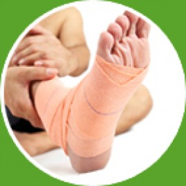
Ankle pain involves any discomfort in one or both ankles.
Causes
Ankle pain is often due to an ankle sprain.
An ankle sprain is an injury to the ligaments, which connect bones to one another.
In most cases, the ankle is twisted inward, causing small tears in the ligaments. The tearing leads to swelling and bruising, making it difficult to bear weight on the joint.
In addition to ankle sprains, ankle pain can be caused by:
Damage or swelling of tendons (which join muscles to bone) or cartilage (which cushions joints)
Infection in the ankle joint
Osteoarthritis, gout,rheumatoid arthritis, Reiter syndrome, and other types of arthritis
Problems in areas near the ankle that can cause you to feel pain in the ankle include:
Blockage of blood vessels in the leg
Heel pain or injuries
Nerve injuries (such as tarsal tunnel syndrome or sciatica)
The elbow is the joint where three long bones meet in the middle portion of the arm. The bone of the upper arm (humerus) meets the inner bone of the forearm (ulna) and the outer bone of the forearm (radius) to form a hinge joint. The radius and ulna also meet in the elbow to allow for rotation of the forearm. The elbow functions to move the arm like a hinge (forward and backward) and in rotation (twisting outward and inward). The biceps muscle is the major muscle that flexes the elbow hinge. The triceps muscle is the major muscle that extends the elbow hinge. The outer bone of the elbow is referred to as the lateral epicondyle and is a part of the humerus bone. Tendons are attached to this area which can be injured, causing inflammation or tendinitis (lateral epicondylitis, or “tennis elbow”).The inner portion of the elbow is a bony prominence called the medial epicondyle. Additional tendons from the muscles attach here and can be injured, causing medial epicondylitis, “golfer’s elbow.” A fluid-filled sac (bursa), which serves to reduce friction, overlies the tip of the elbow (olecranon bursa). The elbow can be affected by inflammation of the tendons or the bursae (plural for bursa) or conditions that affect the bones and joints, such as fractures, arthritis, or nerve irritation. Joint pain in the elbow can result from injury or disease involving any of these structures.
Knee pain
The knee joint consists of an articulation between four bones: the femur, tibia, fibula and patella. There are four compartments to the knee. These are the medial and lateral tibiofemoral compartments, the patellofemoral compartment and the superior tibiofibular joint. The components of each of these compartments can suffer from repetitive strain, injury or disease. Running long distance can cause pain tothe knee joint as it is high impact exercise.
Injuries
Some common injuries include :-
Sprain (Ligament sprain)
Medial collateral ligament
Lateral collateral ligament
Anterior cruciate ligament
Posterior cruciate ligament
Tear of meniscus
Medial meniscus
Lateral meniscus
Strain (Muscle strain)
Quadriceps muscles
Hamstring muscles
Popliteal muscle
Patellar tendon
Hamstring tendon
Popliteal tendon
Hemarthrosis. Hemarthrosis tends to develop over a relatively short period after injury, from several minutes to a few hours.
Diseases
Some of the diseases of cause of knee pain include:
Knee osteoarthritis
Chondromalacia patella
Baker’s cyst
Meniscal cyst
Discoid meniscus
Osgood-Schlatter disease
Larsen-Johansson disease
Knee rheumatoid arthritis
Osteochondritis dissecans disease
Synovial chondromatosis disease
Tumors
Ankylosing spondylitis
Reiter’s syndrome
Tuberculosis arthritis
Septic arthritis (Pyogenic arthritis)
Osteomyelitis
Hemophilic arthritis
Gout (Gouty arthritis)
Inflammations
Bursitis of the knee
Prepatellar bursitis – Housemaid’s knee (most common)
Infrapatellar bursitis – Clergyman’s knee (Superficial infrapatellar bursitis and Deep infrapatellar bursitis)
Semimembranosus bursitis
Tendinitis
Patellar tendinitis (Jumper’s knee)
Hamstring tendinitis
Popliteal tendinitis
Synovitis of the knee
Deformities
Common deformities of the knee include:
Genu varum
Genu valgum
Genu recurvatum (Knee hyperextension)
Knee flexion deformity
Bipartite patella
Syndromes
Patellofemoral pain syndrome (Runner’s knee)
Plica syndrome
Iliotibial band syndrome
Fractures
Femoral fracture
Tibial fracture
Patella fracture
Dislocations
Patella dislocation
Knee joint dislocation (Tibiofemoral joint dislocation)
Referred knee pain
Referred pain is that pain perceived at a site different from its point of origin but innervated by the same spinal segment. Sometimes knee pain may be related to another area from body. For example, knee pain can come from ankle, foot, hip joints or lumbar spine
Ankle pain
Ankle pain involves any discomfort in one or both ankles.
Causes
Ankle pain is often due to an ankle sprain.
An ankle sprain is an injury to the ligaments, which connect bones to one another.
In most cases, the ankle is twisted inward, causing small tears in the ligaments. The tearing leads to swelling and bruising, making it difficult to bear weight on the joint.
In addition to ankle sprains, ankle pain can be caused by:
Damage or swelling of tendons (which join muscles to bone) or cartilage (which cushions joints)
Infection in the ankle joint
Osteoarthritis, gout,rheumatoid arthritis, Reiter syndrome, and other types of arthritis
Problems in areas near the ankle that can cause you to feel pain in the ankle include:
Blockage of blood vessels in the leg
Heel pain or injuries
Nerve injuries (such as tarsal tunnel syndrome or sciatica)
Neck pain (or cervicalgia) is a common problem, with two-thirds of the population having neck painat some point in their lives. Neck pain, although felt in the neck, can be caused by numerous other spinal problems. Neck pain may arise due to muscular tightness in both the neck and upper back, or pinching of the nerves emanating from the cervical vertebrae. Joint disruption in the neck creates pain, as does joint disruption in the upper back.The head is supported by the lower neck and upper back, and it is these areas that commonly cause neck pain.The top three joints in the neck allow for most movement of the neck and head. The lower joints in the neck and those of the upper back create a supportive structure for the head to sit on. If this support system is affected adversely, then the muscles in the area will tighten, leading to neck pain.
Differential diagnosis
Neck pain may come from any of the structures in the neck including: vascular, nerve, airway, digestive, and musculature / skeletal or be referred from other areas of the body.
Major and severe causes of neck pain (roughly in order of severity) include :-
Carotid artery dissection
Referred pain from acute coronary syndrome
Head and neck cancer
Infections: retropharyngeal abscess, epiglottitis, etc.
Spinal disc herniation – protruding or bulging discs, or if severe prolapse.
Spondylosis – degenerative arthritis and osteophytes
Spinal stenosis – a narrowing of the spinal canal
The more common and lesser neck pain causes include :-
Stress – physical and emotional stresses
Prolonged postures – many people fall asleep on sofas and chairs and wake up with sore necks.
Minor injuries and falls – car accidents, sporting events and day to day injuries that are really minor.
Referred pain – mostly from upper back problems
Over-use – muscular strain is one of the most common causes
Whiplash
Herniated disc
Pinched nerv
Differential diagnosis
Neck pain may come from any of the structures in the neck including: vascular, nerve, airway, digestive, and musculature / skeletal or be referred from other areas of the body.
Major and severe causes of neck pain (roughly in order of severity) include :-
Carotid artery dissection
Referred pain from acute coronary syndrome
Head and neck cancer
Infections: retropharyngeal abscess, epiglottitis, etc.
Spinal disc herniation – protruding or bulging discs, or if severe prolapse.
Spondylosis – degenerative arthritis and osteophytes
Spinal stenosis – a narrowing of the spinal canal
The more common and lesser neck pain causes include :-
Stress – physical and emotional stresses
Prolonged postures – many people fall asleep on sofas and chairs and wake up with sore necks.
Minor injuries and falls – car accidents, sporting events and day to day injuries that are really minor.
Referred pain – mostly from upper back problems
Over-use – muscular strain is one of the most common causes
Whiplash
Herniated disc
Pinched nerv
Sports injuries are injuries that occur in athletic activities. They can result from acute trauma, or from overuse of a particular body part.
Classification
Traumatic injuries account for most injuries in contact sports such as Ice Hockey, Association football, rugby league, rugby union, rules football, Gaelic football and football because of the dynamic and high collision nature of these sports, Collisions with the ground, objects, and other players are common, and unexpected dynamic forces on limbs and joints can cause injury.
Traumatic injuries can include :-
Contusion or bruise – damage to small blood vessels which causes bleeding within the tissues.
Strain – trauma to a muscle due to overstreching and tearing of muscle fibers
Sprain – an injury in a joint, caused by the ligament being stretched beyond its own capacity
Wound – abrasion or puncture of the skin
Bone fracture
Head injury
Spinal cord injury
In sports medicine, a catastrophic injury is defined as severe trauma to the human head, spine, or brain.Concussions in sport became a major issue in the United States in the 2000s, as evidence connected repeated concussions and subconcussive hits with chronic traumatic encephalopathy (CTE) and increased suicide risk. It is most pronounced in football, and a related ailment (dementia pugilistica) afflicts boxers, but is also seen in other sports, and in females and adolescents
Overuse and repetitive stress injury problems associated with sports include :-
Runner's knee
Tennis elbow
Tendinosis
Some activities have particular risks; see :-
Bicycle safety
Gun safety
Sailing ship accidents
Skateboarding#Safety
Classification
Traumatic injuries account for most injuries in contact sports such as Ice Hockey, Association football, rugby league, rugby union, rules football, Gaelic football and football because of the dynamic and high collision nature of these sports, Collisions with the ground, objects, and other players are common, and unexpected dynamic forces on limbs and joints can cause injury.
Traumatic injuries can include :-
Contusion or bruise – damage to small blood vessels which causes bleeding within the tissues.
Strain – trauma to a muscle due to overstreching and tearing of muscle fibers
Sprain – an injury in a joint, caused by the ligament being stretched beyond its own capacity
Wound – abrasion or puncture of the skin
Bone fracture
Head injury
Spinal cord injury
In sports medicine, a catastrophic injury is defined as severe trauma to the human head, spine, or brain.Concussions in sport became a major issue in the United States in the 2000s, as evidence connected repeated concussions and subconcussive hits with chronic traumatic encephalopathy (CTE) and increased suicide risk. It is most pronounced in football, and a related ailment (dementia pugilistica) afflicts boxers, but is also seen in other sports, and in females and adolescents
Overuse and repetitive stress injury problems associated with sports include :-
Runner's knee
Tennis elbow
Tendinosis
Some activities have particular risks; see :-
Bicycle safety
Gun safety
Sailing ship accidents
Skateboarding#Safety
Pre-Surgical Education and Rehabilitation
Your physician may refer you physical therapy prior to your scheduled surgery. The purpose of pre-surgical physical therapy will vary. At minimum, your therapist will discuss your post-surgical precautions and/or restrictions. You may be instructed on how to safely move in bed, get in and out of a chair, and use an assistive device, such as crutches ora walker. You may be given exercises to perform at home in order to maximize your range of motion before surgery.Depending on your needs, you may be seen for one visit or you may be seen for multiple visits before your surgery.
Post-Surgical Rehabilitation
Our goal during your post-surgical rehabilitation is to return you to your prior level of physical function. This includes successful return to your work, recreational activities, and competitive sports.Our therapists provide one-on-one treatment sessions to closely monitor your progress and will advance you according to your response as well as your physician’s recommendations. We maintain regular communication with your surgeon to ensure your progress matches the rate of tissue healing so that your recovery is optimal.Your participation in your rehabilitation is critical. On your first post-surgical visit, your therapist will work with you to establish your goals for therapy. Your therapist will review any post-surgical precautions you may have. You will also be prescribed a home exercise program so that you can continue your progress outside of therapy. Commonly treated orthopedic procedures include, but are not limited to:
Rotator cuff repairs
Total joint replacements (shoulder, hip, knee)
Anterior cruciate ligament (ACL) reconstruction
Arthroscopic knee surgeries (such as meniscectomies, meniscal repairs)
Arthroscopic hip surgeries (such as labral repairs)
Achilles tendon repairs
Nerve releases (such as cubital tunnel, radial tunnel, carpal tunnel, tarsal tunnel)
Neck and back surgery (such as fusions, laminectomy, decompression)
Surgical fixation of fractures of the arms and legs
We strongly recommend you schedule your first post-operative visit before you have surgery in order to ensure you are seen in a timely manner. You should ask your physician how soon after surgery you should begin physical therapy
Your physician may refer you physical therapy prior to your scheduled surgery. The purpose of pre-surgical physical therapy will vary. At minimum, your therapist will discuss your post-surgical precautions and/or restrictions. You may be instructed on how to safely move in bed, get in and out of a chair, and use an assistive device, such as crutches ora walker. You may be given exercises to perform at home in order to maximize your range of motion before surgery.Depending on your needs, you may be seen for one visit or you may be seen for multiple visits before your surgery.
Post-Surgical Rehabilitation
Our goal during your post-surgical rehabilitation is to return you to your prior level of physical function. This includes successful return to your work, recreational activities, and competitive sports.Our therapists provide one-on-one treatment sessions to closely monitor your progress and will advance you according to your response as well as your physician’s recommendations. We maintain regular communication with your surgeon to ensure your progress matches the rate of tissue healing so that your recovery is optimal.Your participation in your rehabilitation is critical. On your first post-surgical visit, your therapist will work with you to establish your goals for therapy. Your therapist will review any post-surgical precautions you may have. You will also be prescribed a home exercise program so that you can continue your progress outside of therapy. Commonly treated orthopedic procedures include, but are not limited to:
Rotator cuff repairs
Total joint replacements (shoulder, hip, knee)
Anterior cruciate ligament (ACL) reconstruction
Arthroscopic knee surgeries (such as meniscectomies, meniscal repairs)
Arthroscopic hip surgeries (such as labral repairs)
Achilles tendon repairs
Nerve releases (such as cubital tunnel, radial tunnel, carpal tunnel, tarsal tunnel)
Neck and back surgery (such as fusions, laminectomy, decompression)
Surgical fixation of fractures of the arms and legs
We strongly recommend you schedule your first post-operative visit before you have surgery in order to ensure you are seen in a timely manner. You should ask your physician how soon after surgery you should begin physical therapy
Rehabilitation after stroke, spinal cord injury, brain injury, nerve injury ,poliomylitis, guillain barre syndrome, cerebral palsy, neurological disorder is a continuous process with multidisciplinary approach.
Main Aim of Our Treatments:
Physiotherapeutic Management
Pain Management
Physical Rehabilitation
When most people think of a Physiotherapist they imagine somebody running onto the sports field with a bucket & sponge.While the treatment of sports injuries is one aspect of our profession, Physiotherapy has evolved beyond this & is now acknowledged as an essential element in the management of most musculoskeletal, neurological & rheumatologic conditions.
Our registered Physiotherapists at The Physiotherapy Centre have extensive post-graduate training & vast experience within the NHS, private sector & sports organizations/clubs & offer a wide range of treatment techniques for various conditions including:
Arthritis
Back Pain
Frozen Shoulder
Elbow, Knees ,ankle Pain
Neck & Back Pain
Sports Injuries
Post-Operative Surgery
Joint Replacement
Posture-related Pain
Rheumatoid Arthritis
Repetitive Strain Injury
Fractured Bones
Joint Dislocations
Headaches & Tinnitus
Obstetrics (Pre/Postnatal)
Women’s Health Issues
Nerve Disorders
Stroke
Parkinson’s Disease
Multiple Sclerosis
Treatment may include any or all of these techniques:
DPRC is a leading provider of healthcare & sports injury rehabilitation in Bangladesh and offers unrivalled facilities to help maximize your potential. Our treatment options include:
Electrotherapy (e.g. UST, SWD, MWD, IRR, IFT, TENS, AUTO-TRACTION,WAX BATH, NERVE STIMULAROR-FARADIC/GALVANIC)
Joint Manipulation and Mobilization
Taping/Strapping
Ergonomic/Postural Assessment
Soft Tissue Massage
Gym-based Rehabilitation
Core Stability/Muscle Balance
Joint manipulation or mobilization
Soft tissue massage
Stretches
Supervised exercises
Laser
Specialist Neuro-Physiotherapy
Home exercise program
Physiotherapeutic Management
Pain Management
Physical Rehabilitation
When most people think of a Physiotherapist they imagine somebody running onto the sports field with a bucket & sponge.While the treatment of sports injuries is one aspect of our profession, Physiotherapy has evolved beyond this & is now acknowledged as an essential element in the management of most musculoskeletal, neurological & rheumatologic conditions.
Our registered Physiotherapists at The Physiotherapy Centre have extensive post-graduate training & vast experience within the NHS, private sector & sports organizations/clubs & offer a wide range of treatment techniques for various conditions including:
Arthritis
Back Pain
Frozen Shoulder
Elbow, Knees ,ankle Pain
Neck & Back Pain
Sports Injuries
Post-Operative Surgery
Joint Replacement
Posture-related Pain
Rheumatoid Arthritis
Repetitive Strain Injury
Fractured Bones
Joint Dislocations
Headaches & Tinnitus
Obstetrics (Pre/Postnatal)
Women’s Health Issues
Nerve Disorders
Stroke
Parkinson’s Disease
Multiple Sclerosis
Treatment may include any or all of these techniques:
DPRC is a leading provider of healthcare & sports injury rehabilitation in Bangladesh and offers unrivalled facilities to help maximize your potential. Our treatment options include:
Electrotherapy (e.g. UST, SWD, MWD, IRR, IFT, TENS, AUTO-TRACTION,WAX BATH, NERVE STIMULAROR-FARADIC/GALVANIC)
Joint Manipulation and Mobilization
Taping/Strapping
Ergonomic/Postural Assessment
Soft Tissue Massage
Gym-based Rehabilitation
Core Stability/Muscle Balance
Joint manipulation or mobilization
Soft tissue massage
Stretches
Supervised exercises
Laser
Specialist Neuro-Physiotherapy
Home exercise program
DPRC specialized hospital & research center have a technically & equipmentally most rich Radiology & Imaging department with 128 Slice CT Scan Double unit 1.5T and 18 channel MRI. DPRC specialized hospital & research center Trust applies highest technology to give the best possible diagnosis. Not only Highest quality medical equipments we have the experienced man behind the Machine.
By the combination of high quality Machine, experience manpower, most experienced radiologist, efficient management DPRC specialized hospital & research center rendering world class Radiology & Imaging Services.
openmri
Our radiology and Imaging services are:
MRI
MRI BRAIN
MRCP
MRI & MRA BRAIN
MRI ANKLE (LT./RT.)
MRA NECK
MRI ARM/Forearm
MRI AXILLAE RT./LT.
MRI BRAIN & BASE OF SKULL
MRI BRAIN & CRANIAL NERVES
MRI BRAIN & EAR
MRI BRAIN & ORBIT
MRI BRAIN & PITUITARY GLAND
MRI BRAIN & PNS
MRI BRAIN & SPECTROSCOPY
MR Mammography
CT scan
CT Angiogram
Brain – Head
Pituitary Fossa
Orbits
PNS
Naopharynx
Neck
Chest- Mediastinum
HBS/Upper Abdomen
Whole Abdomen
K.U.B. Region
Lower Abdomen Pelvic Organs
Wholespine
Cervical Spine
Dorsal Spine
Lumbar Spine
Joints
Bones
Mammogram
Both Breasts
Right/left Breast
X-Rays
Chest P/A View
KUB
IVU with post Mict, Film
Retrograde Cystourethroagram
Plain X-Ray Abdomen/C.P.D.
Skull B/V
P.N.S
Cervical Spine B/V
Thoracic Spine B/V
Lumber/Dorso- lumber Spine B/V
Lumbo-Sacral Spine B/V
Other Contrast X-Rays
Ba-Meal stomach & Duodenum
Ba- swallow Oesophagus
Ba-Enema Double Contrrast
Oral Cholecystography
Myelogram
Ultrasound Imaging
GE-Logiq-P5-Premium
Whole Abdomen & Pelvic Organs
HBS/Upper abdomen
Lower abdomen/Uterus & Adnexae
KUB /Genito-Urinary tract
KUB with Prostate & Residual Volume
thyroid
Brain
Breast
Scrotum/Testis
Soft Tissue
Fetal age determination
Routine obstetrical U.S
Color Doppler US of Vertebro-basilar system
Color Doppler US of carotids
Color Doppler US of deep veins of leg
Color Doppler US of Renal Artery
Trans rectal USG
Transvaginal USG
Retro-Urethrogram
Histero-Sulhingogram
Ba-Follow Through
MCU
Ba-Enema
Radiology, Imaging and other Tests
ECG
Holter ECG
ECHO
ETT
Color Doppler
Endoscopy
Colonoscopy
ERCP
BMD
OPG
EEG
By the combination of high quality Machine, experience manpower, most experienced radiologist, efficient management DPRC specialized hospital & research center rendering world class Radiology & Imaging Services.
openmri
Our radiology and Imaging services are:
MRI
MRI BRAIN
MRCP
MRI & MRA BRAIN
MRI ANKLE (LT./RT.)
MRA NECK
MRI ARM/Forearm
MRI AXILLAE RT./LT.
MRI BRAIN & BASE OF SKULL
MRI BRAIN & CRANIAL NERVES
MRI BRAIN & EAR
MRI BRAIN & ORBIT
MRI BRAIN & PITUITARY GLAND
MRI BRAIN & PNS
MRI BRAIN & SPECTROSCOPY
MR Mammography
CT scan
CT Angiogram
Brain – Head
Pituitary Fossa
Orbits
PNS
Naopharynx
Neck
Chest- Mediastinum
HBS/Upper Abdomen
Whole Abdomen
K.U.B. Region
Lower Abdomen Pelvic Organs
Wholespine
Cervical Spine
Dorsal Spine
Lumbar Spine
Joints
Bones
Mammogram
Both Breasts
Right/left Breast
X-Rays
Chest P/A View
KUB
IVU with post Mict, Film
Retrograde Cystourethroagram
Plain X-Ray Abdomen/C.P.D.
Skull B/V
P.N.S
Cervical Spine B/V
Thoracic Spine B/V
Lumber/Dorso- lumber Spine B/V
Lumbo-Sacral Spine B/V
Other Contrast X-Rays
Ba-Meal stomach & Duodenum
Ba- swallow Oesophagus
Ba-Enema Double Contrrast
Oral Cholecystography
Myelogram
Ultrasound Imaging
GE-Logiq-P5-Premium
Whole Abdomen & Pelvic Organs
HBS/Upper abdomen
Lower abdomen/Uterus & Adnexae
KUB /Genito-Urinary tract
KUB with Prostate & Residual Volume
thyroid
Brain
Breast
Scrotum/Testis
Soft Tissue
Fetal age determination
Routine obstetrical U.S
Color Doppler US of Vertebro-basilar system
Color Doppler US of carotids
Color Doppler US of deep veins of leg
Color Doppler US of Renal Artery
Trans rectal USG
Transvaginal USG
Retro-Urethrogram
Histero-Sulhingogram
Ba-Follow Through
MCU
Ba-Enema
Radiology, Imaging and other Tests
ECG
Holter ECG
ECHO
ETT
Color Doppler
Endoscopy
Colonoscopy
ERCP
BMD
OPG
EEG
DPRC specialized hospital & research center believe on quality to ensure world class diagnostic Services. It is one of the largest laboratories in Bangladesh including High-tech Medical equipments and most efficient manpower, and experienced management.
laboratory-ser
Departments of Laboratory Medicine
Biochemistry
Immunology
Hematology
Serology
Microbiology
Histo & cytopathology
Molecular (PCR) Laboratory
We perform about all the investigation which is possible in out country.
Haematological Exam
CBC,ESR
TC,DC,Hb%ESR
CE,Count
Platelet Count
Prothombin Time
Blood Film
Reticulocyte Count
MP
LE Cell
BT,CT
APTT
ASO Titre
R.A. Test
CRP
TPHA
ANF
Anti DNA
Elisa Method Hepatitis (Panel)
Anti HAV-IgM
Anti HBs
Anti HBs
Anti HEV-IgM
HBs Ag
Anti HCV
HBe Ag
Anti HBe
HIV TEST
HIV (1+2)
Hormone Test
T3
FT3
CPK
T4
FT4
TSH
Progesterone
Testosterone
LH
FSH
Cancer Marker
AFP
CEA
BHCG
PSA
Microbiology Exam Blood for C/S throat swab
Gram Stain
C/S
Biochemical Exam
Sugar fasting
S. Random
2 hrs. ABF
After75 gm glucose
S. Bilirubin (Total /Direct/Indireact)
SGT/ALT
Alk,Phosphatase
Acid Phosphatase
S. Cholesterol
HDL Cholesterol
Lipid Profile
Urea/ BUN
S. Electrolytes
S. Total Protein
Globulin
LDH
CPK/CK
GTT
SGOT/AST
S. Amylase
S. Calcium
S. Triglyceride
LDL Cholesterol
S. Phosphate
S. Creatinine
S. Uric Acid
Albumin
AGRatio
YGT
CK-MB
Immunology / Serology
Widal Test
Febrile Antigens
Rose Waaler Test
VDRL
IgE
ANA
Blood group & Rh factor
Rh. Antibody Tire
Coomb’s Test(Direct/Indirect)
Elisa of T-BgA/IgG/IgM/TB
KLB
Sputum
Gram Stain
C/S
Eosinophil
AFB
AFB C/S
laboratory-ser
Departments of Laboratory Medicine
Biochemistry
Immunology
Hematology
Serology
Microbiology
Histo & cytopathology
Molecular (PCR) Laboratory
We perform about all the investigation which is possible in out country.
Haematological Exam
CBC,ESR
TC,DC,Hb%ESR
CE,Count
Platelet Count
Prothombin Time
Blood Film
Reticulocyte Count
MP
LE Cell
BT,CT
APTT
ASO Titre
R.A. Test
CRP
TPHA
ANF
Anti DNA
Elisa Method Hepatitis (Panel)
Anti HAV-IgM
Anti HBs
Anti HBs
Anti HEV-IgM
HBs Ag
Anti HCV
HBe Ag
Anti HBe
HIV TEST
HIV (1+2)
Hormone Test
T3
FT3
CPK
T4
FT4
TSH
Progesterone
Testosterone
LH
FSH
Cancer Marker
AFP
CEA
BHCG
PSA
Microbiology Exam Blood for C/S throat swab
Gram Stain
C/S
Biochemical Exam
Sugar fasting
S. Random
2 hrs. ABF
After75 gm glucose
S. Bilirubin (Total /Direct/Indireact)
SGT/ALT
Alk,Phosphatase
Acid Phosphatase
S. Cholesterol
HDL Cholesterol
Lipid Profile
Urea/ BUN
S. Electrolytes
S. Total Protein
Globulin
LDH
CPK/CK
GTT
SGOT/AST
S. Amylase
S. Calcium
S. Triglyceride
LDL Cholesterol
S. Phosphate
S. Creatinine
S. Uric Acid
Albumin
AGRatio
YGT
CK-MB
Immunology / Serology
Widal Test
Febrile Antigens
Rose Waaler Test
VDRL
IgE
ANA
Blood group & Rh factor
Rh. Antibody Tire
Coomb’s Test(Direct/Indirect)
Elisa of T-BgA/IgG/IgM/TB
KLB
Sputum
Gram Stain
C/S
Eosinophil
AFB
AFB C/S
Pharmacy and orthoses/prostheses
pharmacist-jobs
Arthritis (from Greek arthro-, joint + -itis, inflammation; plural: arthritides) is a form of joint disorder that involves inflammation of one or more joints. There are over 100 different forms of arthritis. The most common form, osteoarthritis (degenerative joint disease), is a result of trauma to the joint, infection of the joint, or age. Other arthritis forms are rheumatoid arthritis, psoriatic arthritis, and related autoimmune diseases. Septic arthritis is caused by joint infection.
The major complaint by individuals who have arthritis is joint pain. Pain is often a constant and may be localized to the joint affected. The pain from arthritis is due to inflammation that occurs around the joint, damage to the joint from disease, daily wear and tear of joint, muscle strains caused by forceful movements against stiff painful joints and fatigue.
The major complaint by individuals who have arthritis is joint pain. Pain is often a constant and may be localized to the joint affected. The pain from arthritis is due to inflammation that occurs around the joint, damage to the joint from disease, daily wear and tear of joint, muscle strains caused by forceful movements against stiff painful joints and fatigue.
Back pain is pain felt in the back that usually originates from the muscles, nerves, bones, joints or other structures in the spine.
Back pain may have a sudden onset or can be a chronic pain; it can be constant or intermittent, stay in one place or radiate to other areas. It may be a dull ache, or a sharp or piercing or burning sensation. The pain may radiate into the arms and hands as well as the legs or feet, and may include symptoms other than pain. These symptoms may include tingling, weakness or numbness.
Back pain is one of humanity’s most frequent complaints. Acute low back pain (also called lumbago) is the fifth most common reason for physician visits. About nine out of ten adults experience back pain at some point in their life, and five out of ten working adults have back pain every year. Back pain is second only to upper respiratory conditions as the stated cause of work loss. Also, it’s the single leading cause of disability worldwide as Americans spend at least $50 billion each year treating it.
The spine is a complex interconnecting network of nerves, joints, muscles, tendons and ligaments, all of which are capable of producing pain. Large nerves that originate in the spine and go to the legs and arms can make pain radiate to the extremities.
Back pain may have a sudden onset or can be a chronic pain; it can be constant or intermittent, stay in one place or radiate to other areas. It may be a dull ache, or a sharp or piercing or burning sensation. The pain may radiate into the arms and hands as well as the legs or feet, and may include symptoms other than pain. These symptoms may include tingling, weakness or numbness.
Back pain is one of humanity’s most frequent complaints. Acute low back pain (also called lumbago) is the fifth most common reason for physician visits. About nine out of ten adults experience back pain at some point in their life, and five out of ten working adults have back pain every year. Back pain is second only to upper respiratory conditions as the stated cause of work loss. Also, it’s the single leading cause of disability worldwide as Americans spend at least $50 billion each year treating it.
The spine is a complex interconnecting network of nerves, joints, muscles, tendons and ligaments, all of which are capable of producing pain. Large nerves that originate in the spine and go to the legs and arms can make pain radiate to the extremities.
Frozen shoulder, medically referred to as adhesive capsulitis, is a disorder in which the shoulder capsule, the connective tissue surrounding the glenohumeral joint of the shoulder, becomes inflamed and stiff, greatly restricting motion and causing chronic pain.
Adhesive capsulitis is a painful and disabling condition that often causes great frustration for patients and caregivers due to slow recovery. Movement of the shoulder is severely restricted. Pain is usually constant, worse at night, and when the weather is colder; and along with the restricted movement can make even small tasks impossible. Certain movements or bumps can cause sudden onset of tremendous pain and cramping that can last several minutes.
This condition, for which an exact cause is unknown, can last from five months to three years or more and is thought in some cases to be caused by injury or trauma to the area. It is believed that it may have an autoimmune component, with the body attacking healthy tissue in the capsule. There is also a lack of fluid in the joint, further restricting movement.
In addition to difficulty with everyday tasks, people who suffer from adhesive capsulitis usually experience problems sleeping for extended periods due to pain that is worse at night and restricted movement/positions. The condition also can lead to depression, pain, and problems in the neck and back.
Risk factors for frozen shoulder include diabetes, stroke, accidents, lung disease, connective tissue disorders, thyroid disease, and heart disease. The condition very rarely appears in people under 40.
Treatment may be painful and taxing and consists of physical therapy, occupational therapy, Chiropractic, medication, massage therapy, hydrodilatation or surgery. A doctor may also perform manipulation under, which breaks up the adhesions and scar tissue in the joint to help restore some range of motion. Pain and inflammation can be controlled with analgesics and NSAIDs. The condition tends to be self-limiting: it usually resolves over time without surgery, but this may take up to two years. Most people regain about 90% of shoulder motion over time. People who suffer from adhesive capsulitis may have extreme difficulty working and going about normal life activities for several months or longer.
Adhesive capsulitis is a painful and disabling condition that often causes great frustration for patients and caregivers due to slow recovery. Movement of the shoulder is severely restricted. Pain is usually constant, worse at night, and when the weather is colder; and along with the restricted movement can make even small tasks impossible. Certain movements or bumps can cause sudden onset of tremendous pain and cramping that can last several minutes.
This condition, for which an exact cause is unknown, can last from five months to three years or more and is thought in some cases to be caused by injury or trauma to the area. It is believed that it may have an autoimmune component, with the body attacking healthy tissue in the capsule. There is also a lack of fluid in the joint, further restricting movement.
In addition to difficulty with everyday tasks, people who suffer from adhesive capsulitis usually experience problems sleeping for extended periods due to pain that is worse at night and restricted movement/positions. The condition also can lead to depression, pain, and problems in the neck and back.
Risk factors for frozen shoulder include diabetes, stroke, accidents, lung disease, connective tissue disorders, thyroid disease, and heart disease. The condition very rarely appears in people under 40.
Treatment may be painful and taxing and consists of physical therapy, occupational therapy, Chiropractic, medication, massage therapy, hydrodilatation or surgery. A doctor may also perform manipulation under, which breaks up the adhesions and scar tissue in the joint to help restore some range of motion. Pain and inflammation can be controlled with analgesics and NSAIDs. The condition tends to be self-limiting: it usually resolves over time without surgery, but this may take up to two years. Most people regain about 90% of shoulder motion over time. People who suffer from adhesive capsulitis may have extreme difficulty working and going about normal life activities for several months or longer.
Elbow pain
The elbow is the joint where three long bones meet in the middle portion of the arm. The bone of the upper arm (humerus) meets the inner bone of the forearm (ulna) and the outer bone of the forearm (radius) to form a hinge joint. The radius and ulna also meet in the elbow to allow for rotation of the forearm. The elbow functions to move the arm like a hinge (forward and backward) and in rotation (twisting outward and inward). The biceps muscle is the major muscle that flexes the elbow hinge. The triceps muscle is the major muscle that extends the elbow hinge. The outer bone of the elbow is referred to as the lateral epicondyle and is a part of the humerus bone. Tendons are attached to this area which can be injured, causing inflammation or tendinitis (lateral epicondylitis, or “tennis elbow”).The inner portion of the elbow is a bony prominence called the medial epicondyle. Additional tendons from the muscles attach here and can be injured, causing medial epicondylitis, “golfer’s elbow.” A fluid-filled sac (bursa), which serves to reduce friction, overlies the tip of the elbow (olecranon bursa). The elbow can be affected by inflammation of the tendons or the bursae (plural for bursa) or conditions that affect the bones and joints, such as fractures, arthritis, or nerve irritation. Joint pain in the elbow can result from injury or disease involving any of these structures.
Knee pain
The knee joint consists of an articulation between four bones: the femur, tibia, fibula and patella. There are four compartments to the knee. These are the medial and lateral tibiofemoral compartments, the patellofemoral compartment and the superior tibiofibular joint. The components of each of these compartments can suffer from repetitive strain, injury or disease. Running long distance can cause pain tothe knee joint as it is high impact exercise.
Injuries
Some common injuries include :-
Sprain (Ligament sprain)
Medial collateral ligament
Lateral collateral ligament
Anterior cruciate ligament
Posterior cruciate ligament
Tear of meniscus
Medial meniscus
Lateral meniscus
Strain (Muscle strain)
Quadriceps muscles
Hamstring muscles
Popliteal muscle
Patellar tendon
Hamstring tendon
Popliteal tendon
Hemarthrosis. Hemarthrosis tends to develop over a relatively short period after injury, from several minutes to a few hours.
Diseases
Some of the diseases of cause of knee pain include:
Knee osteoarthritis
Chondromalacia patella
Baker’s cyst
Meniscal cyst
Discoid meniscus
Osgood-Schlatter disease
Larsen-Johansson disease
Knee rheumatoid arthritis
Osteochondritis dissecans disease
Synovial chondromatosis disease
Tumors
Ankylosing spondylitis
Reiter’s syndrome
Tuberculosis arthritis
Septic arthritis (Pyogenic arthritis)
Osteomyelitis
Hemophilic arthritis
Gout (Gouty arthritis)
Inflammations
Bursitis of the knee
Prepatellar bursitis – Housemaid’s knee (most common)
Infrapatellar bursitis – Clergyman’s knee (Superficial infrapatellar bursitis and Deep infrapatellar bursitis)
Semimembranosus bursitis
Tendinitis
Patellar tendinitis (Jumper’s knee)
Hamstring tendinitis
Popliteal tendinitis
Synovitis of the knee
Deformities
Common deformities of the knee include:
Genu varum
Genu valgum
Genu recurvatum (Knee hyperextension)
Knee flexion deformity
Bipartite patella
Syndromes
Patellofemoral pain syndrome (Runner’s knee)
Plica syndrome
Iliotibial band syndrome
Fractures
Femoral fracture
Tibial fracture
Patella fracture
Dislocations
Patella dislocation
Knee joint dislocation (Tibiofemoral joint dislocation)
Referred knee pain
Referred pain is that pain perceived at a site different from its point of origin but innervated by the same spinal segment. Sometimes knee pain may be related to another area from body. For example, knee pain can come from ankle, foot, hip joints or lumbar spine
Ankle pain
Ankle pain involves any discomfort in one or both ankles.
Causes
Ankle pain is often due to an ankle sprain.
An ankle sprain is an injury to the ligaments, which connect bones to one another.
In most cases, the ankle is twisted inward, causing small tears in the ligaments. The tearing leads to swelling and bruising, making it difficult to bear weight on the joint.
In addition to ankle sprains, ankle pain can be caused by:
Damage or swelling of tendons (which join muscles to bone) or cartilage (which cushions joints)
Infection in the ankle joint
Osteoarthritis, gout,rheumatoid arthritis, Reiter syndrome, and other types of arthritis
Problems in areas near the ankle that can cause you to feel pain in the ankle include:
Blockage of blood vessels in the leg
Heel pain or injuries
Nerve injuries (such as tarsal tunnel syndrome or sciatica)
The elbow is the joint where three long bones meet in the middle portion of the arm. The bone of the upper arm (humerus) meets the inner bone of the forearm (ulna) and the outer bone of the forearm (radius) to form a hinge joint. The radius and ulna also meet in the elbow to allow for rotation of the forearm. The elbow functions to move the arm like a hinge (forward and backward) and in rotation (twisting outward and inward). The biceps muscle is the major muscle that flexes the elbow hinge. The triceps muscle is the major muscle that extends the elbow hinge. The outer bone of the elbow is referred to as the lateral epicondyle and is a part of the humerus bone. Tendons are attached to this area which can be injured, causing inflammation or tendinitis (lateral epicondylitis, or “tennis elbow”).The inner portion of the elbow is a bony prominence called the medial epicondyle. Additional tendons from the muscles attach here and can be injured, causing medial epicondylitis, “golfer’s elbow.” A fluid-filled sac (bursa), which serves to reduce friction, overlies the tip of the elbow (olecranon bursa). The elbow can be affected by inflammation of the tendons or the bursae (plural for bursa) or conditions that affect the bones and joints, such as fractures, arthritis, or nerve irritation. Joint pain in the elbow can result from injury or disease involving any of these structures.
Knee pain
The knee joint consists of an articulation between four bones: the femur, tibia, fibula and patella. There are four compartments to the knee. These are the medial and lateral tibiofemoral compartments, the patellofemoral compartment and the superior tibiofibular joint. The components of each of these compartments can suffer from repetitive strain, injury or disease. Running long distance can cause pain tothe knee joint as it is high impact exercise.
Injuries
Some common injuries include :-
Sprain (Ligament sprain)
Medial collateral ligament
Lateral collateral ligament
Anterior cruciate ligament
Posterior cruciate ligament
Tear of meniscus
Medial meniscus
Lateral meniscus
Strain (Muscle strain)
Quadriceps muscles
Hamstring muscles
Popliteal muscle
Patellar tendon
Hamstring tendon
Popliteal tendon
Hemarthrosis. Hemarthrosis tends to develop over a relatively short period after injury, from several minutes to a few hours.
Diseases
Some of the diseases of cause of knee pain include:
Knee osteoarthritis
Chondromalacia patella
Baker’s cyst
Meniscal cyst
Discoid meniscus
Osgood-Schlatter disease
Larsen-Johansson disease
Knee rheumatoid arthritis
Osteochondritis dissecans disease
Synovial chondromatosis disease
Tumors
Ankylosing spondylitis
Reiter’s syndrome
Tuberculosis arthritis
Septic arthritis (Pyogenic arthritis)
Osteomyelitis
Hemophilic arthritis
Gout (Gouty arthritis)
Inflammations
Bursitis of the knee
Prepatellar bursitis – Housemaid’s knee (most common)
Infrapatellar bursitis – Clergyman’s knee (Superficial infrapatellar bursitis and Deep infrapatellar bursitis)
Semimembranosus bursitis
Tendinitis
Patellar tendinitis (Jumper’s knee)
Hamstring tendinitis
Popliteal tendinitis
Synovitis of the knee
Deformities
Common deformities of the knee include:
Genu varum
Genu valgum
Genu recurvatum (Knee hyperextension)
Knee flexion deformity
Bipartite patella
Syndromes
Patellofemoral pain syndrome (Runner’s knee)
Plica syndrome
Iliotibial band syndrome
Fractures
Femoral fracture
Tibial fracture
Patella fracture
Dislocations
Patella dislocation
Knee joint dislocation (Tibiofemoral joint dislocation)
Referred knee pain
Referred pain is that pain perceived at a site different from its point of origin but innervated by the same spinal segment. Sometimes knee pain may be related to another area from body. For example, knee pain can come from ankle, foot, hip joints or lumbar spine
Ankle pain
Ankle pain involves any discomfort in one or both ankles.
Causes
Ankle pain is often due to an ankle sprain.
An ankle sprain is an injury to the ligaments, which connect bones to one another.
In most cases, the ankle is twisted inward, causing small tears in the ligaments. The tearing leads to swelling and bruising, making it difficult to bear weight on the joint.
In addition to ankle sprains, ankle pain can be caused by:
Damage or swelling of tendons (which join muscles to bone) or cartilage (which cushions joints)
Infection in the ankle joint
Osteoarthritis, gout,rheumatoid arthritis, Reiter syndrome, and other types of arthritis
Problems in areas near the ankle that can cause you to feel pain in the ankle include:
Blockage of blood vessels in the leg
Heel pain or injuries
Nerve injuries (such as tarsal tunnel syndrome or sciatica)
Neck pain (or cervicalgia) is a common problem, with two-thirds of the population having neck painat some point in their lives. Neck pain, although felt in the neck, can be caused by numerous other spinal problems. Neck pain may arise due to muscular tightness in both the neck and upper back, or pinching of the nerves emanating from the cervical vertebrae. Joint disruption in the neck creates pain, as does joint disruption in the upper back.The head is supported by the lower neck and upper back, and it is these areas that commonly cause neck pain.The top three joints in the neck allow for most movement of the neck and head. The lower joints in the neck and those of the upper back create a supportive structure for the head to sit on. If this support system is affected adversely, then the muscles in the area will tighten, leading to neck pain.
Differential diagnosis
Neck pain may come from any of the structures in the neck including: vascular, nerve, airway, digestive, and musculature / skeletal or be referred from other areas of the body.
Major and severe causes of neck pain (roughly in order of severity) include :-
Carotid artery dissection
Referred pain from acute coronary syndrome
Head and neck cancer
Infections: retropharyngeal abscess, epiglottitis, etc.
Spinal disc herniation – protruding or bulging discs, or if severe prolapse.
Spondylosis – degenerative arthritis and osteophytes
Spinal stenosis – a narrowing of the spinal canal
The more common and lesser neck pain causes include :-
Stress – physical and emotional stresses
Prolonged postures – many people fall asleep on sofas and chairs and wake up with sore necks.
Minor injuries and falls – car accidents, sporting events and day to day injuries that are really minor.
Referred pain – mostly from upper back problems
Over-use – muscular strain is one of the most common causes
Whiplash
Herniated disc
Pinched nerv
Differential diagnosis
Neck pain may come from any of the structures in the neck including: vascular, nerve, airway, digestive, and musculature / skeletal or be referred from other areas of the body.
Major and severe causes of neck pain (roughly in order of severity) include :-
Carotid artery dissection
Referred pain from acute coronary syndrome
Head and neck cancer
Infections: retropharyngeal abscess, epiglottitis, etc.
Spinal disc herniation – protruding or bulging discs, or if severe prolapse.
Spondylosis – degenerative arthritis and osteophytes
Spinal stenosis – a narrowing of the spinal canal
The more common and lesser neck pain causes include :-
Stress – physical and emotional stresses
Prolonged postures – many people fall asleep on sofas and chairs and wake up with sore necks.
Minor injuries and falls – car accidents, sporting events and day to day injuries that are really minor.
Referred pain – mostly from upper back problems
Over-use – muscular strain is one of the most common causes
Whiplash
Herniated disc
Pinched nerv
Sports injuries are injuries that occur in athletic activities. They can result from acute trauma, or from overuse of a particular body part.
Classification
Traumatic injuries account for most injuries in contact sports such as Ice Hockey, Association football, rugby league, rugby union, rules football, Gaelic football and football because of the dynamic and high collision nature of these sports, Collisions with the ground, objects, and other players are common, and unexpected dynamic forces on limbs and joints can cause injury.
Traumatic injuries can include :-
Contusion or bruise – damage to small blood vessels which causes bleeding within the tissues.
Strain – trauma to a muscle due to overstreching and tearing of muscle fibers
Sprain – an injury in a joint, caused by the ligament being stretched beyond its own capacity
Wound – abrasion or puncture of the skin
Bone fracture
Head injury
Spinal cord injury
In sports medicine, a catastrophic injury is defined as severe trauma to the human head, spine, or brain.Concussions in sport became a major issue in the United States in the 2000s, as evidence connected repeated concussions and subconcussive hits with chronic traumatic encephalopathy (CTE) and increased suicide risk. It is most pronounced in football, and a related ailment (dementia pugilistica) afflicts boxers, but is also seen in other sports, and in females and adolescents
Overuse and repetitive stress injury problems associated with sports include :-
Runner's knee
Tennis elbow
Tendinosis
Some activities have particular risks; see :-
Bicycle safety
Gun safety
Sailing ship accidents
Skateboarding#Safety
Classification
Traumatic injuries account for most injuries in contact sports such as Ice Hockey, Association football, rugby league, rugby union, rules football, Gaelic football and football because of the dynamic and high collision nature of these sports, Collisions with the ground, objects, and other players are common, and unexpected dynamic forces on limbs and joints can cause injury.
Traumatic injuries can include :-
Contusion or bruise – damage to small blood vessels which causes bleeding within the tissues.
Strain – trauma to a muscle due to overstreching and tearing of muscle fibers
Sprain – an injury in a joint, caused by the ligament being stretched beyond its own capacity
Wound – abrasion or puncture of the skin
Bone fracture
Head injury
Spinal cord injury
In sports medicine, a catastrophic injury is defined as severe trauma to the human head, spine, or brain.Concussions in sport became a major issue in the United States in the 2000s, as evidence connected repeated concussions and subconcussive hits with chronic traumatic encephalopathy (CTE) and increased suicide risk. It is most pronounced in football, and a related ailment (dementia pugilistica) afflicts boxers, but is also seen in other sports, and in females and adolescents
Overuse and repetitive stress injury problems associated with sports include :-
Runner's knee
Tennis elbow
Tendinosis
Some activities have particular risks; see :-
Bicycle safety
Gun safety
Sailing ship accidents
Skateboarding#Safety
Pre-Surgical Education and Rehabilitation
Your physician may refer you physical therapy prior to your scheduled surgery. The purpose of pre-surgical physical therapy will vary. At minimum, your therapist will discuss your post-surgical precautions and/or restrictions. You may be instructed on how to safely move in bed, get in and out of a chair, and use an assistive device, such as crutches ora walker. You may be given exercises to perform at home in order to maximize your range of motion before surgery.Depending on your needs, you may be seen for one visit or you may be seen for multiple visits before your surgery.
Post-Surgical Rehabilitation
Our goal during your post-surgical rehabilitation is to return you to your prior level of physical function. This includes successful return to your work, recreational activities, and competitive sports.Our therapists provide one-on-one treatment sessions to closely monitor your progress and will advance you according to your response as well as your physician’s recommendations. We maintain regular communication with your surgeon to ensure your progress matches the rate of tissue healing so that your recovery is optimal.Your participation in your rehabilitation is critical. On your first post-surgical visit, your therapist will work with you to establish your goals for therapy. Your therapist will review any post-surgical precautions you may have. You will also be prescribed a home exercise program so that you can continue your progress outside of therapy. Commonly treated orthopedic procedures include, but are not limited to:
Rotator cuff repairs
Total joint replacements (shoulder, hip, knee)
Anterior cruciate ligament (ACL) reconstruction
Arthroscopic knee surgeries (such as meniscectomies, meniscal repairs)
Arthroscopic hip surgeries (such as labral repairs)
Achilles tendon repairs
Nerve releases (such as cubital tunnel, radial tunnel, carpal tunnel, tarsal tunnel)
Neck and back surgery (such as fusions, laminectomy, decompression)
Surgical fixation of fractures of the arms and legs
We strongly recommend you schedule your first post-operative visit before you have surgery in order to ensure you are seen in a timely manner. You should ask your physician how soon after surgery you should begin physical therapy
Your physician may refer you physical therapy prior to your scheduled surgery. The purpose of pre-surgical physical therapy will vary. At minimum, your therapist will discuss your post-surgical precautions and/or restrictions. You may be instructed on how to safely move in bed, get in and out of a chair, and use an assistive device, such as crutches ora walker. You may be given exercises to perform at home in order to maximize your range of motion before surgery.Depending on your needs, you may be seen for one visit or you may be seen for multiple visits before your surgery.
Post-Surgical Rehabilitation
Our goal during your post-surgical rehabilitation is to return you to your prior level of physical function. This includes successful return to your work, recreational activities, and competitive sports.Our therapists provide one-on-one treatment sessions to closely monitor your progress and will advance you according to your response as well as your physician’s recommendations. We maintain regular communication with your surgeon to ensure your progress matches the rate of tissue healing so that your recovery is optimal.Your participation in your rehabilitation is critical. On your first post-surgical visit, your therapist will work with you to establish your goals for therapy. Your therapist will review any post-surgical precautions you may have. You will also be prescribed a home exercise program so that you can continue your progress outside of therapy. Commonly treated orthopedic procedures include, but are not limited to:
Rotator cuff repairs
Total joint replacements (shoulder, hip, knee)
Anterior cruciate ligament (ACL) reconstruction
Arthroscopic knee surgeries (such as meniscectomies, meniscal repairs)
Arthroscopic hip surgeries (such as labral repairs)
Achilles tendon repairs
Nerve releases (such as cubital tunnel, radial tunnel, carpal tunnel, tarsal tunnel)
Neck and back surgery (such as fusions, laminectomy, decompression)
Surgical fixation of fractures of the arms and legs
We strongly recommend you schedule your first post-operative visit before you have surgery in order to ensure you are seen in a timely manner. You should ask your physician how soon after surgery you should begin physical therapy
Rehabilitation after stroke, spinal cord injury, brain injury, nerve injury ,poliomylitis, guillain barre syndrome, cerebral palsy, neurological disorder is a continuous process with multidisciplinary approach.
Main Aim of Our Treatments:
Physiotherapeutic Management
Pain Management
Physical Rehabilitation
When most people think of a Physiotherapist they imagine somebody running onto the sports field with a bucket & sponge.While the treatment of sports injuries is one aspect of our profession, Physiotherapy has evolved beyond this & is now acknowledged as an essential element in the management of most musculoskeletal, neurological & rheumatologic conditions.
Our registered Physiotherapists at The Physiotherapy Centre have extensive post-graduate training & vast experience within the NHS, private sector & sports organizations/clubs & offer a wide range of treatment techniques for various conditions including:
Arthritis
Back Pain
Frozen Shoulder
Elbow, Knees ,ankle Pain
Neck & Back Pain
Sports Injuries
Post-Operative Surgery
Joint Replacement
Posture-related Pain
Rheumatoid Arthritis
Repetitive Strain Injury
Fractured Bones
Joint Dislocations
Headaches & Tinnitus
Obstetrics (Pre/Postnatal)
Women’s Health Issues
Nerve Disorders
Stroke
Parkinson’s Disease
Multiple Sclerosis
Treatment may include any or all of these techniques:
DPRC is a leading provider of healthcare & sports injury rehabilitation in Bangladesh and offers unrivalled facilities to help maximize your potential. Our treatment options include:
Electrotherapy (e.g. UST, SWD, MWD, IRR, IFT, TENS, AUTO-TRACTION,WAX BATH, NERVE STIMULAROR-FARADIC/GALVANIC)
Joint Manipulation and Mobilization
Taping/Strapping
Ergonomic/Postural Assessment
Soft Tissue Massage
Gym-based Rehabilitation
Core Stability/Muscle Balance
Joint manipulation or mobilization
Soft tissue massage
Stretches
Supervised exercises
Laser
Specialist Neuro-Physiotherapy
Home exercise program
Physiotherapeutic Management
Pain Management
Physical Rehabilitation
When most people think of a Physiotherapist they imagine somebody running onto the sports field with a bucket & sponge.While the treatment of sports injuries is one aspect of our profession, Physiotherapy has evolved beyond this & is now acknowledged as an essential element in the management of most musculoskeletal, neurological & rheumatologic conditions.
Our registered Physiotherapists at The Physiotherapy Centre have extensive post-graduate training & vast experience within the NHS, private sector & sports organizations/clubs & offer a wide range of treatment techniques for various conditions including:
Arthritis
Back Pain
Frozen Shoulder
Elbow, Knees ,ankle Pain
Neck & Back Pain
Sports Injuries
Post-Operative Surgery
Joint Replacement
Posture-related Pain
Rheumatoid Arthritis
Repetitive Strain Injury
Fractured Bones
Joint Dislocations
Headaches & Tinnitus
Obstetrics (Pre/Postnatal)
Women’s Health Issues
Nerve Disorders
Stroke
Parkinson’s Disease
Multiple Sclerosis
Treatment may include any or all of these techniques:
DPRC is a leading provider of healthcare & sports injury rehabilitation in Bangladesh and offers unrivalled facilities to help maximize your potential. Our treatment options include:
Electrotherapy (e.g. UST, SWD, MWD, IRR, IFT, TENS, AUTO-TRACTION,WAX BATH, NERVE STIMULAROR-FARADIC/GALVANIC)
Joint Manipulation and Mobilization
Taping/Strapping
Ergonomic/Postural Assessment
Soft Tissue Massage
Gym-based Rehabilitation
Core Stability/Muscle Balance
Joint manipulation or mobilization
Soft tissue massage
Stretches
Supervised exercises
Laser
Specialist Neuro-Physiotherapy
Home exercise program
DPRC specialized hospital & research center have a technically & equipmentally most rich Radiology & Imaging department with 128 Slice CT Scan Double unit 1.5T and 18 channel MRI. DPRC specialized hospital & research center Trust applies highest technology to give the best possible diagnosis. Not only Highest quality medical equipments we have the experienced man behind the Machine.
By the combination of high quality Machine, experience manpower, most experienced radiologist, efficient management DPRC specialized hospital & research center rendering world class Radiology & Imaging Services.
openmri
Our radiology and Imaging services are:
MRI
MRI BRAIN
MRCP
MRI & MRA BRAIN
MRI ANKLE (LT./RT.)
MRA NECK
MRI ARM/Forearm
MRI AXILLAE RT./LT.
MRI BRAIN & BASE OF SKULL
MRI BRAIN & CRANIAL NERVES
MRI BRAIN & EAR
MRI BRAIN & ORBIT
MRI BRAIN & PITUITARY GLAND
MRI BRAIN & PNS
MRI BRAIN & SPECTROSCOPY
MR Mammography
CT scan
CT Angiogram
Brain – Head
Pituitary Fossa
Orbits
PNS
Naopharynx
Neck
Chest- Mediastinum
HBS/Upper Abdomen
Whole Abdomen
K.U.B. Region
Lower Abdomen Pelvic Organs
Wholespine
Cervical Spine
Dorsal Spine
Lumbar Spine
Joints
Bones
Mammogram
Both Breasts
Right/left Breast
X-Rays
Chest P/A View
KUB
IVU with post Mict, Film
Retrograde Cystourethroagram
Plain X-Ray Abdomen/C.P.D.
Skull B/V
P.N.S
Cervical Spine B/V
Thoracic Spine B/V
Lumber/Dorso- lumber Spine B/V
Lumbo-Sacral Spine B/V
Other Contrast X-Rays
Ba-Meal stomach & Duodenum
Ba- swallow Oesophagus
Ba-Enema Double Contrrast
Oral Cholecystography
Myelogram
Ultrasound Imaging
GE-Logiq-P5-Premium
Whole Abdomen & Pelvic Organs
HBS/Upper abdomen
Lower abdomen/Uterus & Adnexae
KUB /Genito-Urinary tract
KUB with Prostate & Residual Volume
thyroid
Brain
Breast
Scrotum/Testis
Soft Tissue
Fetal age determination
Routine obstetrical U.S
Color Doppler US of Vertebro-basilar system
Color Doppler US of carotids
Color Doppler US of deep veins of leg
Color Doppler US of Renal Artery
Trans rectal USG
Transvaginal USG
Retro-Urethrogram
Histero-Sulhingogram
Ba-Follow Through
MCU
Ba-Enema
Radiology, Imaging and other Tests
ECG
Holter ECG
ECHO
ETT
Color Doppler
Endoscopy
Colonoscopy
ERCP
BMD
OPG
EEG
By the combination of high quality Machine, experience manpower, most experienced radiologist, efficient management DPRC specialized hospital & research center rendering world class Radiology & Imaging Services.
openmri
Our radiology and Imaging services are:
MRI
MRI BRAIN
MRCP
MRI & MRA BRAIN
MRI ANKLE (LT./RT.)
MRA NECK
MRI ARM/Forearm
MRI AXILLAE RT./LT.
MRI BRAIN & BASE OF SKULL
MRI BRAIN & CRANIAL NERVES
MRI BRAIN & EAR
MRI BRAIN & ORBIT
MRI BRAIN & PITUITARY GLAND
MRI BRAIN & PNS
MRI BRAIN & SPECTROSCOPY
MR Mammography
CT scan
CT Angiogram
Brain – Head
Pituitary Fossa
Orbits
PNS
Naopharynx
Neck
Chest- Mediastinum
HBS/Upper Abdomen
Whole Abdomen
K.U.B. Region
Lower Abdomen Pelvic Organs
Wholespine
Cervical Spine
Dorsal Spine
Lumbar Spine
Joints
Bones
Mammogram
Both Breasts
Right/left Breast
X-Rays
Chest P/A View
KUB
IVU with post Mict, Film
Retrograde Cystourethroagram
Plain X-Ray Abdomen/C.P.D.
Skull B/V
P.N.S
Cervical Spine B/V
Thoracic Spine B/V
Lumber/Dorso- lumber Spine B/V
Lumbo-Sacral Spine B/V
Other Contrast X-Rays
Ba-Meal stomach & Duodenum
Ba- swallow Oesophagus
Ba-Enema Double Contrrast
Oral Cholecystography
Myelogram
Ultrasound Imaging
GE-Logiq-P5-Premium
Whole Abdomen & Pelvic Organs
HBS/Upper abdomen
Lower abdomen/Uterus & Adnexae
KUB /Genito-Urinary tract
KUB with Prostate & Residual Volume
thyroid
Brain
Breast
Scrotum/Testis
Soft Tissue
Fetal age determination
Routine obstetrical U.S
Color Doppler US of Vertebro-basilar system
Color Doppler US of carotids
Color Doppler US of deep veins of leg
Color Doppler US of Renal Artery
Trans rectal USG
Transvaginal USG
Retro-Urethrogram
Histero-Sulhingogram
Ba-Follow Through
MCU
Ba-Enema
Radiology, Imaging and other Tests
ECG
Holter ECG
ECHO
ETT
Color Doppler
Endoscopy
Colonoscopy
ERCP
BMD
OPG
EEG
DPRC specialized hospital & research center believe on quality to ensure world class diagnostic Services. It is one of the largest laboratories in Bangladesh including High-tech Medical equipments and most efficient manpower, and experienced management.
laboratory-ser
Departments of Laboratory Medicine
Biochemistry
Immunology
Hematology
Serology
Microbiology
Histo & cytopathology
Molecular (PCR) Laboratory
We perform about all the investigation which is possible in out country.
Haematological Exam
CBC,ESR
TC,DC,Hb%ESR
CE,Count
Platelet Count
Prothombin Time
Blood Film
Reticulocyte Count
MP
LE Cell
BT,CT
APTT
ASO Titre
R.A. Test
CRP
TPHA
ANF
Anti DNA
Elisa Method Hepatitis (Panel)
Anti HAV-IgM
Anti HBs
Anti HBs
Anti HEV-IgM
HBs Ag
Anti HCV
HBe Ag
Anti HBe
HIV TEST
HIV (1+2)
Hormone Test
T3
FT3
CPK
T4
FT4
TSH
Progesterone
Testosterone
LH
FSH
Cancer Marker
AFP
CEA
BHCG
PSA
Microbiology Exam Blood for C/S throat swab
Gram Stain
C/S
Biochemical Exam
Sugar fasting
S. Random
2 hrs. ABF
After75 gm glucose
S. Bilirubin (Total /Direct/Indireact)
SGT/ALT
Alk,Phosphatase
Acid Phosphatase
S. Cholesterol
HDL Cholesterol
Lipid Profile
Urea/ BUN
S. Electrolytes
S. Total Protein
Globulin
LDH
CPK/CK
GTT
SGOT/AST
S. Amylase
S. Calcium
S. Triglyceride
LDL Cholesterol
S. Phosphate
S. Creatinine
S. Uric Acid
Albumin
AGRatio
YGT
CK-MB
Immunology / Serology
Widal Test
Febrile Antigens
Rose Waaler Test
VDRL
IgE
ANA
Blood group & Rh factor
Rh. Antibody Tire
Coomb’s Test(Direct/Indirect)
Elisa of T-BgA/IgG/IgM/TB
KLB
Sputum
Gram Stain
C/S
Eosinophil
AFB
AFB C/S
laboratory-ser
Departments of Laboratory Medicine
Biochemistry
Immunology
Hematology
Serology
Microbiology
Histo & cytopathology
Molecular (PCR) Laboratory
We perform about all the investigation which is possible in out country.
Haematological Exam
CBC,ESR
TC,DC,Hb%ESR
CE,Count
Platelet Count
Prothombin Time
Blood Film
Reticulocyte Count
MP
LE Cell
BT,CT
APTT
ASO Titre
R.A. Test
CRP
TPHA
ANF
Anti DNA
Elisa Method Hepatitis (Panel)
Anti HAV-IgM
Anti HBs
Anti HBs
Anti HEV-IgM
HBs Ag
Anti HCV
HBe Ag
Anti HBe
HIV TEST
HIV (1+2)
Hormone Test
T3
FT3
CPK
T4
FT4
TSH
Progesterone
Testosterone
LH
FSH
Cancer Marker
AFP
CEA
BHCG
PSA
Microbiology Exam Blood for C/S throat swab
Gram Stain
C/S
Biochemical Exam
Sugar fasting
S. Random
2 hrs. ABF
After75 gm glucose
S. Bilirubin (Total /Direct/Indireact)
SGT/ALT
Alk,Phosphatase
Acid Phosphatase
S. Cholesterol
HDL Cholesterol
Lipid Profile
Urea/ BUN
S. Electrolytes
S. Total Protein
Globulin
LDH
CPK/CK
GTT
SGOT/AST
S. Amylase
S. Calcium
S. Triglyceride
LDL Cholesterol
S. Phosphate
S. Creatinine
S. Uric Acid
Albumin
AGRatio
YGT
CK-MB
Immunology / Serology
Widal Test
Febrile Antigens
Rose Waaler Test
VDRL
IgE
ANA
Blood group & Rh factor
Rh. Antibody Tire
Coomb’s Test(Direct/Indirect)
Elisa of T-BgA/IgG/IgM/TB
KLB
Sputum
Gram Stain
C/S
Eosinophil
AFB
AFB C/S
Pharmacy and orthoses/prostheses
pharmacist-jobs
Arthritis (from Greek arthro-, joint + -itis, inflammation; plural: arthritides) is a form of joint disorder that involves inflammation of one or more joints. There are over 100 different forms of arthritis. The most common form, osteoarthritis (degenerative joint disease), is a result of trauma to the joint, infection of the joint, or age. Other arthritis forms are rheumatoid arthritis, psoriatic arthritis, and related autoimmune diseases. Septic arthritis is caused by joint infection.
The major complaint by individuals who have arthritis is joint pain. Pain is often a constant and may be localized to the joint affected. The pain from arthritis is due to inflammation that occurs around the joint, damage to the joint from disease, daily wear and tear of joint, muscle strains caused by forceful movements against stiff painful joints and fatigue.
The major complaint by individuals who have arthritis is joint pain. Pain is often a constant and may be localized to the joint affected. The pain from arthritis is due to inflammation that occurs around the joint, damage to the joint from disease, daily wear and tear of joint, muscle strains caused by forceful movements against stiff painful joints and fatigue.
Back pain is pain felt in the back that usually originates from the muscles, nerves, bones, joints or other structures in the spine.
Back pain may have a sudden onset or can be a chronic pain; it can be constant or intermittent, stay in one place or radiate to other areas. It may be a dull ache, or a sharp or piercing or burning sensation. The pain may radiate into the arms and hands as well as the legs or feet, and may include symptoms other than pain. These symptoms may include tingling, weakness or numbness.
Back pain is one of humanity’s most frequent complaints. Acute low back pain (also called lumbago) is the fifth most common reason for physician visits. About nine out of ten adults experience back pain at some point in their life, and five out of ten working adults have back pain every year. Back pain is second only to upper respiratory conditions as the stated cause of work loss. Also, it’s the single leading cause of disability worldwide as Americans spend at least $50 billion each year treating it.
The spine is a complex interconnecting network of nerves, joints, muscles, tendons and ligaments, all of which are capable of producing pain. Large nerves that originate in the spine and go to the legs and arms can make pain radiate to the extremities.
Back pain may have a sudden onset or can be a chronic pain; it can be constant or intermittent, stay in one place or radiate to other areas. It may be a dull ache, or a sharp or piercing or burning sensation. The pain may radiate into the arms and hands as well as the legs or feet, and may include symptoms other than pain. These symptoms may include tingling, weakness or numbness.
Back pain is one of humanity’s most frequent complaints. Acute low back pain (also called lumbago) is the fifth most common reason for physician visits. About nine out of ten adults experience back pain at some point in their life, and five out of ten working adults have back pain every year. Back pain is second only to upper respiratory conditions as the stated cause of work loss. Also, it’s the single leading cause of disability worldwide as Americans spend at least $50 billion each year treating it.
The spine is a complex interconnecting network of nerves, joints, muscles, tendons and ligaments, all of which are capable of producing pain. Large nerves that originate in the spine and go to the legs and arms can make pain radiate to the extremities.
Frozen shoulder, medically referred to as adhesive capsulitis, is a disorder in which the shoulder capsule, the connective tissue surrounding the glenohumeral joint of the shoulder, becomes inflamed and stiff, greatly restricting motion and causing chronic pain.
Adhesive capsulitis is a painful and disabling condition that often causes great frustration for patients and caregivers due to slow recovery. Movement of the shoulder is severely restricted. Pain is usually constant, worse at night, and when the weather is colder; and along with the restricted movement can make even small tasks impossible. Certain movements or bumps can cause sudden onset of tremendous pain and cramping that can last several minutes.
This condition, for which an exact cause is unknown, can last from five months to three years or more and is thought in some cases to be caused by injury or trauma to the area. It is believed that it may have an autoimmune component, with the body attacking healthy tissue in the capsule. There is also a lack of fluid in the joint, further restricting movement.
In addition to difficulty with everyday tasks, people who suffer from adhesive capsulitis usually experience problems sleeping for extended periods due to pain that is worse at night and restricted movement/positions. The condition also can lead to depression, pain, and problems in the neck and back.
Risk factors for frozen shoulder include diabetes, stroke, accidents, lung disease, connective tissue disorders, thyroid disease, and heart disease. The condition very rarely appears in people under 40.
Treatment may be painful and taxing and consists of physical therapy, occupational therapy, Chiropractic, medication, massage therapy, hydrodilatation or surgery. A doctor may also perform manipulation under, which breaks up the adhesions and scar tissue in the joint to help restore some range of motion. Pain and inflammation can be controlled with analgesics and NSAIDs. The condition tends to be self-limiting: it usually resolves over time without surgery, but this may take up to two years. Most people regain about 90% of shoulder motion over time. People who suffer from adhesive capsulitis may have extreme difficulty working and going about normal life activities for several months or longer.
Adhesive capsulitis is a painful and disabling condition that often causes great frustration for patients and caregivers due to slow recovery. Movement of the shoulder is severely restricted. Pain is usually constant, worse at night, and when the weather is colder; and along with the restricted movement can make even small tasks impossible. Certain movements or bumps can cause sudden onset of tremendous pain and cramping that can last several minutes.
This condition, for which an exact cause is unknown, can last from five months to three years or more and is thought in some cases to be caused by injury or trauma to the area. It is believed that it may have an autoimmune component, with the body attacking healthy tissue in the capsule. There is also a lack of fluid in the joint, further restricting movement.
In addition to difficulty with everyday tasks, people who suffer from adhesive capsulitis usually experience problems sleeping for extended periods due to pain that is worse at night and restricted movement/positions. The condition also can lead to depression, pain, and problems in the neck and back.
Risk factors for frozen shoulder include diabetes, stroke, accidents, lung disease, connective tissue disorders, thyroid disease, and heart disease. The condition very rarely appears in people under 40.
Treatment may be painful and taxing and consists of physical therapy, occupational therapy, Chiropractic, medication, massage therapy, hydrodilatation or surgery. A doctor may also perform manipulation under, which breaks up the adhesions and scar tissue in the joint to help restore some range of motion. Pain and inflammation can be controlled with analgesics and NSAIDs. The condition tends to be self-limiting: it usually resolves over time without surgery, but this may take up to two years. Most people regain about 90% of shoulder motion over time. People who suffer from adhesive capsulitis may have extreme difficulty working and going about normal life activities for several months or longer.
Elbow pain
The elbow is the joint where three long bones meet in the middle portion of the arm. The bone of the upper arm (humerus) meets the inner bone of the forearm (ulna) and the outer bone of the forearm (radius) to form a hinge joint. The radius and ulna also meet in the elbow to allow for rotation of the forearm. The elbow functions to move the arm like a hinge (forward and backward) and in rotation (twisting outward and inward). The biceps muscle is the major muscle that flexes the elbow hinge. The triceps muscle is the major muscle that extends the elbow hinge. The outer bone of the elbow is referred to as the lateral epicondyle and is a part of the humerus bone. Tendons are attached to this area which can be injured, causing inflammation or tendinitis (lateral epicondylitis, or “tennis elbow”).The inner portion of the elbow is a bony prominence called the medial epicondyle. Additional tendons from the muscles attach here and can be injured, causing medial epicondylitis, “golfer’s elbow.” A fluid-filled sac (bursa), which serves to reduce friction, overlies the tip of the elbow (olecranon bursa). The elbow can be affected by inflammation of the tendons or the bursae (plural for bursa) or conditions that affect the bones and joints, such as fractures, arthritis, or nerve irritation. Joint pain in the elbow can result from injury or disease involving any of these structures.
Knee pain
The knee joint consists of an articulation between four bones: the femur, tibia, fibula and patella. There are four compartments to the knee. These are the medial and lateral tibiofemoral compartments, the patellofemoral compartment and the superior tibiofibular joint. The components of each of these compartments can suffer from repetitive strain, injury or disease. Running long distance can cause pain tothe knee joint as it is high impact exercise.
Injuries
Some common injuries include :-
Sprain (Ligament sprain)
Medial collateral ligament
Lateral collateral ligament
Anterior cruciate ligament
Posterior cruciate ligament
Tear of meniscus
Medial meniscus
Lateral meniscus
Strain (Muscle strain)
Quadriceps muscles
Hamstring muscles
Popliteal muscle
Patellar tendon
Hamstring tendon
Popliteal tendon
Hemarthrosis. Hemarthrosis tends to develop over a relatively short period after injury, from several minutes to a few hours.
Diseases
Some of the diseases of cause of knee pain include:
Knee osteoarthritis
Chondromalacia patella
Baker’s cyst
Meniscal cyst
Discoid meniscus
Osgood-Schlatter disease
Larsen-Johansson disease
Knee rheumatoid arthritis
Osteochondritis dissecans disease
Synovial chondromatosis disease
Tumors
Ankylosing spondylitis
Reiter’s syndrome
Tuberculosis arthritis
Septic arthritis (Pyogenic arthritis)
Osteomyelitis
Hemophilic arthritis
Gout (Gouty arthritis)
Inflammations
Bursitis of the knee
Prepatellar bursitis – Housemaid’s knee (most common)
Infrapatellar bursitis – Clergyman’s knee (Superficial infrapatellar bursitis and Deep infrapatellar bursitis)
Semimembranosus bursitis
Tendinitis
Patellar tendinitis (Jumper’s knee)
Hamstring tendinitis
Popliteal tendinitis
Synovitis of the knee
Deformities
Common deformities of the knee include:
Genu varum
Genu valgum
Genu recurvatum (Knee hyperextension)
Knee flexion deformity
Bipartite patella
Syndromes
Patellofemoral pain syndrome (Runner’s knee)
Plica syndrome
Iliotibial band syndrome
Fractures
Femoral fracture
Tibial fracture
Patella fracture
Dislocations
Patella dislocation
Knee joint dislocation (Tibiofemoral joint dislocation)
Referred knee pain
Referred pain is that pain perceived at a site different from its point of origin but innervated by the same spinal segment. Sometimes knee pain may be related to another area from body. For example, knee pain can come from ankle, foot, hip joints or lumbar spine
Ankle pain
Ankle pain involves any discomfort in one or both ankles.
Causes
Ankle pain is often due to an ankle sprain.
An ankle sprain is an injury to the ligaments, which connect bones to one another.
In most cases, the ankle is twisted inward, causing small tears in the ligaments. The tearing leads to swelling and bruising, making it difficult to bear weight on the joint.
In addition to ankle sprains, ankle pain can be caused by:
Damage or swelling of tendons (which join muscles to bone) or cartilage (which cushions joints)
Infection in the ankle joint
Osteoarthritis, gout,rheumatoid arthritis, Reiter syndrome, and other types of arthritis
Problems in areas near the ankle that can cause you to feel pain in the ankle include:
Blockage of blood vessels in the leg
Heel pain or injuries
Nerve injuries (such as tarsal tunnel syndrome or sciatica)
The elbow is the joint where three long bones meet in the middle portion of the arm. The bone of the upper arm (humerus) meets the inner bone of the forearm (ulna) and the outer bone of the forearm (radius) to form a hinge joint. The radius and ulna also meet in the elbow to allow for rotation of the forearm. The elbow functions to move the arm like a hinge (forward and backward) and in rotation (twisting outward and inward). The biceps muscle is the major muscle that flexes the elbow hinge. The triceps muscle is the major muscle that extends the elbow hinge. The outer bone of the elbow is referred to as the lateral epicondyle and is a part of the humerus bone. Tendons are attached to this area which can be injured, causing inflammation or tendinitis (lateral epicondylitis, or “tennis elbow”).The inner portion of the elbow is a bony prominence called the medial epicondyle. Additional tendons from the muscles attach here and can be injured, causing medial epicondylitis, “golfer’s elbow.” A fluid-filled sac (bursa), which serves to reduce friction, overlies the tip of the elbow (olecranon bursa). The elbow can be affected by inflammation of the tendons or the bursae (plural for bursa) or conditions that affect the bones and joints, such as fractures, arthritis, or nerve irritation. Joint pain in the elbow can result from injury or disease involving any of these structures.
Knee pain
The knee joint consists of an articulation between four bones: the femur, tibia, fibula and patella. There are four compartments to the knee. These are the medial and lateral tibiofemoral compartments, the patellofemoral compartment and the superior tibiofibular joint. The components of each of these compartments can suffer from repetitive strain, injury or disease. Running long distance can cause pain tothe knee joint as it is high impact exercise.
Injuries
Some common injuries include :-
Sprain (Ligament sprain)
Medial collateral ligament
Lateral collateral ligament
Anterior cruciate ligament
Posterior cruciate ligament
Tear of meniscus
Medial meniscus
Lateral meniscus
Strain (Muscle strain)
Quadriceps muscles
Hamstring muscles
Popliteal muscle
Patellar tendon
Hamstring tendon
Popliteal tendon
Hemarthrosis. Hemarthrosis tends to develop over a relatively short period after injury, from several minutes to a few hours.
Diseases
Some of the diseases of cause of knee pain include:
Knee osteoarthritis
Chondromalacia patella
Baker’s cyst
Meniscal cyst
Discoid meniscus
Osgood-Schlatter disease
Larsen-Johansson disease
Knee rheumatoid arthritis
Osteochondritis dissecans disease
Synovial chondromatosis disease
Tumors
Ankylosing spondylitis
Reiter’s syndrome
Tuberculosis arthritis
Septic arthritis (Pyogenic arthritis)
Osteomyelitis
Hemophilic arthritis
Gout (Gouty arthritis)
Inflammations
Bursitis of the knee
Prepatellar bursitis – Housemaid’s knee (most common)
Infrapatellar bursitis – Clergyman’s knee (Superficial infrapatellar bursitis and Deep infrapatellar bursitis)
Semimembranosus bursitis
Tendinitis
Patellar tendinitis (Jumper’s knee)
Hamstring tendinitis
Popliteal tendinitis
Synovitis of the knee
Deformities
Common deformities of the knee include:
Genu varum
Genu valgum
Genu recurvatum (Knee hyperextension)
Knee flexion deformity
Bipartite patella
Syndromes
Patellofemoral pain syndrome (Runner’s knee)
Plica syndrome
Iliotibial band syndrome
Fractures
Femoral fracture
Tibial fracture
Patella fracture
Dislocations
Patella dislocation
Knee joint dislocation (Tibiofemoral joint dislocation)
Referred knee pain
Referred pain is that pain perceived at a site different from its point of origin but innervated by the same spinal segment. Sometimes knee pain may be related to another area from body. For example, knee pain can come from ankle, foot, hip joints or lumbar spine
Ankle pain
Ankle pain involves any discomfort in one or both ankles.
Causes
Ankle pain is often due to an ankle sprain.
An ankle sprain is an injury to the ligaments, which connect bones to one another.
In most cases, the ankle is twisted inward, causing small tears in the ligaments. The tearing leads to swelling and bruising, making it difficult to bear weight on the joint.
In addition to ankle sprains, ankle pain can be caused by:
Damage or swelling of tendons (which join muscles to bone) or cartilage (which cushions joints)
Infection in the ankle joint
Osteoarthritis, gout,rheumatoid arthritis, Reiter syndrome, and other types of arthritis
Problems in areas near the ankle that can cause you to feel pain in the ankle include:
Blockage of blood vessels in the leg
Heel pain or injuries
Nerve injuries (such as tarsal tunnel syndrome or sciatica)
Neck pain (or cervicalgia) is a common problem, with two-thirds of the population having neck painat some point in their lives. Neck pain, although felt in the neck, can be caused by numerous other spinal problems. Neck pain may arise due to muscular tightness in both the neck and upper back, or pinching of the nerves emanating from the cervical vertebrae. Joint disruption in the neck creates pain, as does joint disruption in the upper back.The head is supported by the lower neck and upper back, and it is these areas that commonly cause neck pain.The top three joints in the neck allow for most movement of the neck and head. The lower joints in the neck and those of the upper back create a supportive structure for the head to sit on. If this support system is affected adversely, then the muscles in the area will tighten, leading to neck pain.
Differential diagnosis
Neck pain may come from any of the structures in the neck including: vascular, nerve, airway, digestive, and musculature / skeletal or be referred from other areas of the body.
Major and severe causes of neck pain (roughly in order of severity) include :-
Carotid artery dissection
Referred pain from acute coronary syndrome
Head and neck cancer
Infections: retropharyngeal abscess, epiglottitis, etc.
Spinal disc herniation – protruding or bulging discs, or if severe prolapse.
Spondylosis – degenerative arthritis and osteophytes
Spinal stenosis – a narrowing of the spinal canal
The more common and lesser neck pain causes include :-
Stress – physical and emotional stresses
Prolonged postures – many people fall asleep on sofas and chairs and wake up with sore necks.
Minor injuries and falls – car accidents, sporting events and day to day injuries that are really minor.
Referred pain – mostly from upper back problems
Over-use – muscular strain is one of the most common causes
Whiplash
Herniated disc
Pinched nerv
Differential diagnosis
Neck pain may come from any of the structures in the neck including: vascular, nerve, airway, digestive, and musculature / skeletal or be referred from other areas of the body.
Major and severe causes of neck pain (roughly in order of severity) include :-
Carotid artery dissection
Referred pain from acute coronary syndrome
Head and neck cancer
Infections: retropharyngeal abscess, epiglottitis, etc.
Spinal disc herniation – protruding or bulging discs, or if severe prolapse.
Spondylosis – degenerative arthritis and osteophytes
Spinal stenosis – a narrowing of the spinal canal
The more common and lesser neck pain causes include :-
Stress – physical and emotional stresses
Prolonged postures – many people fall asleep on sofas and chairs and wake up with sore necks.
Minor injuries and falls – car accidents, sporting events and day to day injuries that are really minor.
Referred pain – mostly from upper back problems
Over-use – muscular strain is one of the most common causes
Whiplash
Herniated disc
Pinched nerv
Sports injuries are injuries that occur in athletic activities. They can result from acute trauma, or from overuse of a particular body part.
Classification
Traumatic injuries account for most injuries in contact sports such as Ice Hockey, Association football, rugby league, rugby union, rules football, Gaelic football and football because of the dynamic and high collision nature of these sports, Collisions with the ground, objects, and other players are common, and unexpected dynamic forces on limbs and joints can cause injury.
Traumatic injuries can include :-
Contusion or bruise – damage to small blood vessels which causes bleeding within the tissues.
Strain – trauma to a muscle due to overstreching and tearing of muscle fibers
Sprain – an injury in a joint, caused by the ligament being stretched beyond its own capacity
Wound – abrasion or puncture of the skin
Bone fracture
Head injury
Spinal cord injury
In sports medicine, a catastrophic injury is defined as severe trauma to the human head, spine, or brain.Concussions in sport became a major issue in the United States in the 2000s, as evidence connected repeated concussions and subconcussive hits with chronic traumatic encephalopathy (CTE) and increased suicide risk. It is most pronounced in football, and a related ailment (dementia pugilistica) afflicts boxers, but is also seen in other sports, and in females and adolescents
Overuse and repetitive stress injury problems associated with sports include :-
Runner's knee
Tennis elbow
Tendinosis
Some activities have particular risks; see :-
Bicycle safety
Gun safety
Sailing ship accidents
Skateboarding#Safety
Classification
Traumatic injuries account for most injuries in contact sports such as Ice Hockey, Association football, rugby league, rugby union, rules football, Gaelic football and football because of the dynamic and high collision nature of these sports, Collisions with the ground, objects, and other players are common, and unexpected dynamic forces on limbs and joints can cause injury.
Traumatic injuries can include :-
Contusion or bruise – damage to small blood vessels which causes bleeding within the tissues.
Strain – trauma to a muscle due to overstreching and tearing of muscle fibers
Sprain – an injury in a joint, caused by the ligament being stretched beyond its own capacity
Wound – abrasion or puncture of the skin
Bone fracture
Head injury
Spinal cord injury
In sports medicine, a catastrophic injury is defined as severe trauma to the human head, spine, or brain.Concussions in sport became a major issue in the United States in the 2000s, as evidence connected repeated concussions and subconcussive hits with chronic traumatic encephalopathy (CTE) and increased suicide risk. It is most pronounced in football, and a related ailment (dementia pugilistica) afflicts boxers, but is also seen in other sports, and in females and adolescents
Overuse and repetitive stress injury problems associated with sports include :-
Runner's knee
Tennis elbow
Tendinosis
Some activities have particular risks; see :-
Bicycle safety
Gun safety
Sailing ship accidents
Skateboarding#Safety
Pre-Surgical Education and Rehabilitation
Your physician may refer you physical therapy prior to your scheduled surgery. The purpose of pre-surgical physical therapy will vary. At minimum, your therapist will discuss your post-surgical precautions and/or restrictions. You may be instructed on how to safely move in bed, get in and out of a chair, and use an assistive device, such as crutches ora walker. You may be given exercises to perform at home in order to maximize your range of motion before surgery.Depending on your needs, you may be seen for one visit or you may be seen for multiple visits before your surgery.
Post-Surgical Rehabilitation
Our goal during your post-surgical rehabilitation is to return you to your prior level of physical function. This includes successful return to your work, recreational activities, and competitive sports.Our therapists provide one-on-one treatment sessions to closely monitor your progress and will advance you according to your response as well as your physician’s recommendations. We maintain regular communication with your surgeon to ensure your progress matches the rate of tissue healing so that your recovery is optimal.Your participation in your rehabilitation is critical. On your first post-surgical visit, your therapist will work with you to establish your goals for therapy. Your therapist will review any post-surgical precautions you may have. You will also be prescribed a home exercise program so that you can continue your progress outside of therapy. Commonly treated orthopedic procedures include, but are not limited to:
Rotator cuff repairs
Total joint replacements (shoulder, hip, knee)
Anterior cruciate ligament (ACL) reconstruction
Arthroscopic knee surgeries (such as meniscectomies, meniscal repairs)
Arthroscopic hip surgeries (such as labral repairs)
Achilles tendon repairs
Nerve releases (such as cubital tunnel, radial tunnel, carpal tunnel, tarsal tunnel)
Neck and back surgery (such as fusions, laminectomy, decompression)
Surgical fixation of fractures of the arms and legs
We strongly recommend you schedule your first post-operative visit before you have surgery in order to ensure you are seen in a timely manner. You should ask your physician how soon after surgery you should begin physical therapy
Your physician may refer you physical therapy prior to your scheduled surgery. The purpose of pre-surgical physical therapy will vary. At minimum, your therapist will discuss your post-surgical precautions and/or restrictions. You may be instructed on how to safely move in bed, get in and out of a chair, and use an assistive device, such as crutches ora walker. You may be given exercises to perform at home in order to maximize your range of motion before surgery.Depending on your needs, you may be seen for one visit or you may be seen for multiple visits before your surgery.
Post-Surgical Rehabilitation
Our goal during your post-surgical rehabilitation is to return you to your prior level of physical function. This includes successful return to your work, recreational activities, and competitive sports.Our therapists provide one-on-one treatment sessions to closely monitor your progress and will advance you according to your response as well as your physician’s recommendations. We maintain regular communication with your surgeon to ensure your progress matches the rate of tissue healing so that your recovery is optimal.Your participation in your rehabilitation is critical. On your first post-surgical visit, your therapist will work with you to establish your goals for therapy. Your therapist will review any post-surgical precautions you may have. You will also be prescribed a home exercise program so that you can continue your progress outside of therapy. Commonly treated orthopedic procedures include, but are not limited to:
Rotator cuff repairs
Total joint replacements (shoulder, hip, knee)
Anterior cruciate ligament (ACL) reconstruction
Arthroscopic knee surgeries (such as meniscectomies, meniscal repairs)
Arthroscopic hip surgeries (such as labral repairs)
Achilles tendon repairs
Nerve releases (such as cubital tunnel, radial tunnel, carpal tunnel, tarsal tunnel)
Neck and back surgery (such as fusions, laminectomy, decompression)
Surgical fixation of fractures of the arms and legs
We strongly recommend you schedule your first post-operative visit before you have surgery in order to ensure you are seen in a timely manner. You should ask your physician how soon after surgery you should begin physical therapy
Rehabilitation after stroke, spinal cord injury, brain injury, nerve injury ,poliomylitis, guillain barre syndrome, cerebral palsy, neurological disorder is a continuous process with multidisciplinary approach.
Main Aim of Our Treatments:
Physiotherapeutic Management
Pain Management
Physical Rehabilitation
When most people think of a Physiotherapist they imagine somebody running onto the sports field with a bucket & sponge.While the treatment of sports injuries is one aspect of our profession, Physiotherapy has evolved beyond this & is now acknowledged as an essential element in the management of most musculoskeletal, neurological & rheumatologic conditions.
Our registered Physiotherapists at The Physiotherapy Centre have extensive post-graduate training & vast experience within the NHS, private sector & sports organizations/clubs & offer a wide range of treatment techniques for various conditions including:
Arthritis
Back Pain
Frozen Shoulder
Elbow, Knees ,ankle Pain
Neck & Back Pain
Sports Injuries
Post-Operative Surgery
Joint Replacement
Posture-related Pain
Rheumatoid Arthritis
Repetitive Strain Injury
Fractured Bones
Joint Dislocations
Headaches & Tinnitus
Obstetrics (Pre/Postnatal)
Women’s Health Issues
Nerve Disorders
Stroke
Parkinson’s Disease
Multiple Sclerosis
Treatment may include any or all of these techniques:
DPRC is a leading provider of healthcare & sports injury rehabilitation in Bangladesh and offers unrivalled facilities to help maximize your potential. Our treatment options include:
Electrotherapy (e.g. UST, SWD, MWD, IRR, IFT, TENS, AUTO-TRACTION,WAX BATH, NERVE STIMULAROR-FARADIC/GALVANIC)
Joint Manipulation and Mobilization
Taping/Strapping
Ergonomic/Postural Assessment
Soft Tissue Massage
Gym-based Rehabilitation
Core Stability/Muscle Balance
Joint manipulation or mobilization
Soft tissue massage
Stretches
Supervised exercises
Laser
Specialist Neuro-Physiotherapy
Home exercise program
Physiotherapeutic Management
Pain Management
Physical Rehabilitation
When most people think of a Physiotherapist they imagine somebody running onto the sports field with a bucket & sponge.While the treatment of sports injuries is one aspect of our profession, Physiotherapy has evolved beyond this & is now acknowledged as an essential element in the management of most musculoskeletal, neurological & rheumatologic conditions.
Our registered Physiotherapists at The Physiotherapy Centre have extensive post-graduate training & vast experience within the NHS, private sector & sports organizations/clubs & offer a wide range of treatment techniques for various conditions including:
Arthritis
Back Pain
Frozen Shoulder
Elbow, Knees ,ankle Pain
Neck & Back Pain
Sports Injuries
Post-Operative Surgery
Joint Replacement
Posture-related Pain
Rheumatoid Arthritis
Repetitive Strain Injury
Fractured Bones
Joint Dislocations
Headaches & Tinnitus
Obstetrics (Pre/Postnatal)
Women’s Health Issues
Nerve Disorders
Stroke
Parkinson’s Disease
Multiple Sclerosis
Treatment may include any or all of these techniques:
DPRC is a leading provider of healthcare & sports injury rehabilitation in Bangladesh and offers unrivalled facilities to help maximize your potential. Our treatment options include:
Electrotherapy (e.g. UST, SWD, MWD, IRR, IFT, TENS, AUTO-TRACTION,WAX BATH, NERVE STIMULAROR-FARADIC/GALVANIC)
Joint Manipulation and Mobilization
Taping/Strapping
Ergonomic/Postural Assessment
Soft Tissue Massage
Gym-based Rehabilitation
Core Stability/Muscle Balance
Joint manipulation or mobilization
Soft tissue massage
Stretches
Supervised exercises
Laser
Specialist Neuro-Physiotherapy
Home exercise program
DPRC specialized hospital & research center have a technically & equipmentally most rich Radiology & Imaging department with 128 Slice CT Scan Double unit 1.5T and 18 channel MRI. DPRC specialized hospital & research center Trust applies highest technology to give the best possible diagnosis. Not only Highest quality medical equipments we have the experienced man behind the Machine.
By the combination of high quality Machine, experience manpower, most experienced radiologist, efficient management DPRC specialized hospital & research center rendering world class Radiology & Imaging Services.
openmri
Our radiology and Imaging services are:
MRI
MRI BRAIN
MRCP
MRI & MRA BRAIN
MRI ANKLE (LT./RT.)
MRA NECK
MRI ARM/Forearm
MRI AXILLAE RT./LT.
MRI BRAIN & BASE OF SKULL
MRI BRAIN & CRANIAL NERVES
MRI BRAIN & EAR
MRI BRAIN & ORBIT
MRI BRAIN & PITUITARY GLAND
MRI BRAIN & PNS
MRI BRAIN & SPECTROSCOPY
MR Mammography
CT scan
CT Angiogram
Brain – Head
Pituitary Fossa
Orbits
PNS
Naopharynx
Neck
Chest- Mediastinum
HBS/Upper Abdomen
Whole Abdomen
K.U.B. Region
Lower Abdomen Pelvic Organs
Wholespine
Cervical Spine
Dorsal Spine
Lumbar Spine
Joints
Bones
Mammogram
Both Breasts
Right/left Breast
X-Rays
Chest P/A View
KUB
IVU with post Mict, Film
Retrograde Cystourethroagram
Plain X-Ray Abdomen/C.P.D.
Skull B/V
P.N.S
Cervical Spine B/V
Thoracic Spine B/V
Lumber/Dorso- lumber Spine B/V
Lumbo-Sacral Spine B/V
Other Contrast X-Rays
Ba-Meal stomach & Duodenum
Ba- swallow Oesophagus
Ba-Enema Double Contrrast
Oral Cholecystography
Myelogram
Ultrasound Imaging
GE-Logiq-P5-Premium
Whole Abdomen & Pelvic Organs
HBS/Upper abdomen
Lower abdomen/Uterus & Adnexae
KUB /Genito-Urinary tract
KUB with Prostate & Residual Volume
thyroid
Brain
Breast
Scrotum/Testis
Soft Tissue
Fetal age determination
Routine obstetrical U.S
Color Doppler US of Vertebro-basilar system
Color Doppler US of carotids
Color Doppler US of deep veins of leg
Color Doppler US of Renal Artery
Trans rectal USG
Transvaginal USG
Retro-Urethrogram
Histero-Sulhingogram
Ba-Follow Through
MCU
Ba-Enema
Radiology, Imaging and other Tests
ECG
Holter ECG
ECHO
ETT
Color Doppler
Endoscopy
Colonoscopy
ERCP
BMD
OPG
EEG
By the combination of high quality Machine, experience manpower, most experienced radiologist, efficient management DPRC specialized hospital & research center rendering world class Radiology & Imaging Services.
openmri
Our radiology and Imaging services are:
MRI
MRI BRAIN
MRCP
MRI & MRA BRAIN
MRI ANKLE (LT./RT.)
MRA NECK
MRI ARM/Forearm
MRI AXILLAE RT./LT.
MRI BRAIN & BASE OF SKULL
MRI BRAIN & CRANIAL NERVES
MRI BRAIN & EAR
MRI BRAIN & ORBIT
MRI BRAIN & PITUITARY GLAND
MRI BRAIN & PNS
MRI BRAIN & SPECTROSCOPY
MR Mammography
CT scan
CT Angiogram
Brain – Head
Pituitary Fossa
Orbits
PNS
Naopharynx
Neck
Chest- Mediastinum
HBS/Upper Abdomen
Whole Abdomen
K.U.B. Region
Lower Abdomen Pelvic Organs
Wholespine
Cervical Spine
Dorsal Spine
Lumbar Spine
Joints
Bones
Mammogram
Both Breasts
Right/left Breast
X-Rays
Chest P/A View
KUB
IVU with post Mict, Film
Retrograde Cystourethroagram
Plain X-Ray Abdomen/C.P.D.
Skull B/V
P.N.S
Cervical Spine B/V
Thoracic Spine B/V
Lumber/Dorso- lumber Spine B/V
Lumbo-Sacral Spine B/V
Other Contrast X-Rays
Ba-Meal stomach & Duodenum
Ba- swallow Oesophagus
Ba-Enema Double Contrrast
Oral Cholecystography
Myelogram
Ultrasound Imaging
GE-Logiq-P5-Premium
Whole Abdomen & Pelvic Organs
HBS/Upper abdomen
Lower abdomen/Uterus & Adnexae
KUB /Genito-Urinary tract
KUB with Prostate & Residual Volume
thyroid
Brain
Breast
Scrotum/Testis
Soft Tissue
Fetal age determination
Routine obstetrical U.S
Color Doppler US of Vertebro-basilar system
Color Doppler US of carotids
Color Doppler US of deep veins of leg
Color Doppler US of Renal Artery
Trans rectal USG
Transvaginal USG
Retro-Urethrogram
Histero-Sulhingogram
Ba-Follow Through
MCU
Ba-Enema
Radiology, Imaging and other Tests
ECG
Holter ECG
ECHO
ETT
Color Doppler
Endoscopy
Colonoscopy
ERCP
BMD
OPG
EEG
DPRC specialized hospital & research center believe on quality to ensure world class diagnostic Services. It is one of the largest laboratories in Bangladesh including High-tech Medical equipments and most efficient manpower, and experienced management.
laboratory-ser
Departments of Laboratory Medicine
Biochemistry
Immunology
Hematology
Serology
Microbiology
Histo & cytopathology
Molecular (PCR) Laboratory
We perform about all the investigation which is possible in out country.
Haematological Exam
CBC,ESR
TC,DC,Hb%ESR
CE,Count
Platelet Count
Prothombin Time
Blood Film
Reticulocyte Count
MP
LE Cell
BT,CT
APTT
ASO Titre
R.A. Test
CRP
TPHA
ANF
Anti DNA
Elisa Method Hepatitis (Panel)
Anti HAV-IgM
Anti HBs
Anti HBs
Anti HEV-IgM
HBs Ag
Anti HCV
HBe Ag
Anti HBe
HIV TEST
HIV (1+2)
Hormone Test
T3
FT3
CPK
T4
FT4
TSH
Progesterone
Testosterone
LH
FSH
Cancer Marker
AFP
CEA
BHCG
PSA
Microbiology Exam Blood for C/S throat swab
Gram Stain
C/S
Biochemical Exam
Sugar fasting
S. Random
2 hrs. ABF
After75 gm glucose
S. Bilirubin (Total /Direct/Indireact)
SGT/ALT
Alk,Phosphatase
Acid Phosphatase
S. Cholesterol
HDL Cholesterol
Lipid Profile
Urea/ BUN
S. Electrolytes
S. Total Protein
Globulin
LDH
CPK/CK
GTT
SGOT/AST
S. Amylase
S. Calcium
S. Triglyceride
LDL Cholesterol
S. Phosphate
S. Creatinine
S. Uric Acid
Albumin
AGRatio
YGT
CK-MB
Immunology / Serology
Widal Test
Febrile Antigens
Rose Waaler Test
VDRL
IgE
ANA
Blood group & Rh factor
Rh. Antibody Tire
Coomb’s Test(Direct/Indirect)
Elisa of T-BgA/IgG/IgM/TB
KLB
Sputum
Gram Stain
C/S
Eosinophil
AFB
AFB C/S
laboratory-ser
Departments of Laboratory Medicine
Biochemistry
Immunology
Hematology
Serology
Microbiology
Histo & cytopathology
Molecular (PCR) Laboratory
We perform about all the investigation which is possible in out country.
Haematological Exam
CBC,ESR
TC,DC,Hb%ESR
CE,Count
Platelet Count
Prothombin Time
Blood Film
Reticulocyte Count
MP
LE Cell
BT,CT
APTT
ASO Titre
R.A. Test
CRP
TPHA
ANF
Anti DNA
Elisa Method Hepatitis (Panel)
Anti HAV-IgM
Anti HBs
Anti HBs
Anti HEV-IgM
HBs Ag
Anti HCV
HBe Ag
Anti HBe
HIV TEST
HIV (1+2)
Hormone Test
T3
FT3
CPK
T4
FT4
TSH
Progesterone
Testosterone
LH
FSH
Cancer Marker
AFP
CEA
BHCG
PSA
Microbiology Exam Blood for C/S throat swab
Gram Stain
C/S
Biochemical Exam
Sugar fasting
S. Random
2 hrs. ABF
After75 gm glucose
S. Bilirubin (Total /Direct/Indireact)
SGT/ALT
Alk,Phosphatase
Acid Phosphatase
S. Cholesterol
HDL Cholesterol
Lipid Profile
Urea/ BUN
S. Electrolytes
S. Total Protein
Globulin
LDH
CPK/CK
GTT
SGOT/AST
S. Amylase
S. Calcium
S. Triglyceride
LDL Cholesterol
S. Phosphate
S. Creatinine
S. Uric Acid
Albumin
AGRatio
YGT
CK-MB
Immunology / Serology
Widal Test
Febrile Antigens
Rose Waaler Test
VDRL
IgE
ANA
Blood group & Rh factor
Rh. Antibody Tire
Coomb’s Test(Direct/Indirect)
Elisa of T-BgA/IgG/IgM/TB
KLB
Sputum
Gram Stain
C/S
Eosinophil
AFB
AFB C/S
Pharmacy and orthoses/prostheses
pharmacist-jobs
Arthritis (from Greek arthro-, joint + -itis, inflammation; plural: arthritides) is a form of joint disorder that involves inflammation of one or more joints. There are over 100 different forms of arthritis. The most common form, osteoarthritis (degenerative joint disease), is a result of trauma to the joint, infection of the joint, or age. Other arthritis forms are rheumatoid arthritis, psoriatic arthritis, and related autoimmune diseases. Septic arthritis is caused by joint infection.
The major complaint by individuals who have arthritis is joint pain. Pain is often a constant and may be localized to the joint affected. The pain from arthritis is due to inflammation that occurs around the joint, damage to the joint from disease, daily wear and tear of joint, muscle strains caused by forceful movements against stiff painful joints and fatigue.
The major complaint by individuals who have arthritis is joint pain. Pain is often a constant and may be localized to the joint affected. The pain from arthritis is due to inflammation that occurs around the joint, damage to the joint from disease, daily wear and tear of joint, muscle strains caused by forceful movements against stiff painful joints and fatigue.
Back pain is pain felt in the back that usually originates from the muscles, nerves, bones, joints or other structures in the spine.
Back pain may have a sudden onset or can be a chronic pain; it can be constant or intermittent, stay in one place or radiate to other areas. It may be a dull ache, or a sharp or piercing or burning sensation. The pain may radiate into the arms and hands as well as the legs or feet, and may include symptoms other than pain. These symptoms may include tingling, weakness or numbness.
Back pain is one of humanity’s most frequent complaints. Acute low back pain (also called lumbago) is the fifth most common reason for physician visits. About nine out of ten adults experience back pain at some point in their life, and five out of ten working adults have back pain every year. Back pain is second only to upper respiratory conditions as the stated cause of work loss. Also, it’s the single leading cause of disability worldwide as Americans spend at least $50 billion each year treating it.
The spine is a complex interconnecting network of nerves, joints, muscles, tendons and ligaments, all of which are capable of producing pain. Large nerves that originate in the spine and go to the legs and arms can make pain radiate to the extremities.
Back pain may have a sudden onset or can be a chronic pain; it can be constant or intermittent, stay in one place or radiate to other areas. It may be a dull ache, or a sharp or piercing or burning sensation. The pain may radiate into the arms and hands as well as the legs or feet, and may include symptoms other than pain. These symptoms may include tingling, weakness or numbness.
Back pain is one of humanity’s most frequent complaints. Acute low back pain (also called lumbago) is the fifth most common reason for physician visits. About nine out of ten adults experience back pain at some point in their life, and five out of ten working adults have back pain every year. Back pain is second only to upper respiratory conditions as the stated cause of work loss. Also, it’s the single leading cause of disability worldwide as Americans spend at least $50 billion each year treating it.
The spine is a complex interconnecting network of nerves, joints, muscles, tendons and ligaments, all of which are capable of producing pain. Large nerves that originate in the spine and go to the legs and arms can make pain radiate to the extremities.
Frozen shoulder, medically referred to as adhesive capsulitis, is a disorder in which the shoulder capsule, the connective tissue surrounding the glenohumeral joint of the shoulder, becomes inflamed and stiff, greatly restricting motion and causing chronic pain.
Adhesive capsulitis is a painful and disabling condition that often causes great frustration for patients and caregivers due to slow recovery. Movement of the shoulder is severely restricted. Pain is usually constant, worse at night, and when the weather is colder; and along with the restricted movement can make even small tasks impossible. Certain movements or bumps can cause sudden onset of tremendous pain and cramping that can last several minutes.
This condition, for which an exact cause is unknown, can last from five months to three years or more and is thought in some cases to be caused by injury or trauma to the area. It is believed that it may have an autoimmune component, with the body attacking healthy tissue in the capsule. There is also a lack of fluid in the joint, further restricting movement.
In addition to difficulty with everyday tasks, people who suffer from adhesive capsulitis usually experience problems sleeping for extended periods due to pain that is worse at night and restricted movement/positions. The condition also can lead to depression, pain, and problems in the neck and back.
Risk factors for frozen shoulder include diabetes, stroke, accidents, lung disease, connective tissue disorders, thyroid disease, and heart disease. The condition very rarely appears in people under 40.
Treatment may be painful and taxing and consists of physical therapy, occupational therapy, Chiropractic, medication, massage therapy, hydrodilatation or surgery. A doctor may also perform manipulation under, which breaks up the adhesions and scar tissue in the joint to help restore some range of motion. Pain and inflammation can be controlled with analgesics and NSAIDs. The condition tends to be self-limiting: it usually resolves over time without surgery, but this may take up to two years. Most people regain about 90% of shoulder motion over time. People who suffer from adhesive capsulitis may have extreme difficulty working and going about normal life activities for several months or longer.
Adhesive capsulitis is a painful and disabling condition that often causes great frustration for patients and caregivers due to slow recovery. Movement of the shoulder is severely restricted. Pain is usually constant, worse at night, and when the weather is colder; and along with the restricted movement can make even small tasks impossible. Certain movements or bumps can cause sudden onset of tremendous pain and cramping that can last several minutes.
This condition, for which an exact cause is unknown, can last from five months to three years or more and is thought in some cases to be caused by injury or trauma to the area. It is believed that it may have an autoimmune component, with the body attacking healthy tissue in the capsule. There is also a lack of fluid in the joint, further restricting movement.
In addition to difficulty with everyday tasks, people who suffer from adhesive capsulitis usually experience problems sleeping for extended periods due to pain that is worse at night and restricted movement/positions. The condition also can lead to depression, pain, and problems in the neck and back.
Risk factors for frozen shoulder include diabetes, stroke, accidents, lung disease, connective tissue disorders, thyroid disease, and heart disease. The condition very rarely appears in people under 40.
Treatment may be painful and taxing and consists of physical therapy, occupational therapy, Chiropractic, medication, massage therapy, hydrodilatation or surgery. A doctor may also perform manipulation under, which breaks up the adhesions and scar tissue in the joint to help restore some range of motion. Pain and inflammation can be controlled with analgesics and NSAIDs. The condition tends to be self-limiting: it usually resolves over time without surgery, but this may take up to two years. Most people regain about 90% of shoulder motion over time. People who suffer from adhesive capsulitis may have extreme difficulty working and going about normal life activities for several months or longer.
Elbow pain
The elbow is the joint where three long bones meet in the middle portion of the arm. The bone of the upper arm (humerus) meets the inner bone of the forearm (ulna) and the outer bone of the forearm (radius) to form a hinge joint. The radius and ulna also meet in the elbow to allow for rotation of the forearm. The elbow functions to move the arm like a hinge (forward and backward) and in rotation (twisting outward and inward). The biceps muscle is the major muscle that flexes the elbow hinge. The triceps muscle is the major muscle that extends the elbow hinge. The outer bone of the elbow is referred to as the lateral epicondyle and is a part of the humerus bone. Tendons are attached to this area which can be injured, causing inflammation or tendinitis (lateral epicondylitis, or “tennis elbow”).The inner portion of the elbow is a bony prominence called the medial epicondyle. Additional tendons from the muscles attach here and can be injured, causing medial epicondylitis, “golfer’s elbow.” A fluid-filled sac (bursa), which serves to reduce friction, overlies the tip of the elbow (olecranon bursa). The elbow can be affected by inflammation of the tendons or the bursae (plural for bursa) or conditions that affect the bones and joints, such as fractures, arthritis, or nerve irritation. Joint pain in the elbow can result from injury or disease involving any of these structures.
Knee pain
The knee joint consists of an articulation between four bones: the femur, tibia, fibula and patella. There are four compartments to the knee. These are the medial and lateral tibiofemoral compartments, the patellofemoral compartment and the superior tibiofibular joint. The components of each of these compartments can suffer from repetitive strain, injury or disease. Running long distance can cause pain tothe knee joint as it is high impact exercise.
Injuries
Some common injuries include :-
Sprain (Ligament sprain)
Medial collateral ligament
Lateral collateral ligament
Anterior cruciate ligament
Posterior cruciate ligament
Tear of meniscus
Medial meniscus
Lateral meniscus
Strain (Muscle strain)
Quadriceps muscles
Hamstring muscles
Popliteal muscle
Patellar tendon
Hamstring tendon
Popliteal tendon
Hemarthrosis. Hemarthrosis tends to develop over a relatively short period after injury, from several minutes to a few hours.
Diseases
Some of the diseases of cause of knee pain include:
Knee osteoarthritis
Chondromalacia patella
Baker’s cyst
Meniscal cyst
Discoid meniscus
Osgood-Schlatter disease
Larsen-Johansson disease
Knee rheumatoid arthritis
Osteochondritis dissecans disease
Synovial chondromatosis disease
Tumors
Ankylosing spondylitis
Reiter’s syndrome
Tuberculosis arthritis
Septic arthritis (Pyogenic arthritis)
Osteomyelitis
Hemophilic arthritis
Gout (Gouty arthritis)
Inflammations
Bursitis of the knee
Prepatellar bursitis – Housemaid’s knee (most common)
Infrapatellar bursitis – Clergyman’s knee (Superficial infrapatellar bursitis and Deep infrapatellar bursitis)
Semimembranosus bursitis
Tendinitis
Patellar tendinitis (Jumper’s knee)
Hamstring tendinitis
Popliteal tendinitis
Synovitis of the knee
Deformities
Common deformities of the knee include:
Genu varum
Genu valgum
Genu recurvatum (Knee hyperextension)
Knee flexion deformity
Bipartite patella
Syndromes
Patellofemoral pain syndrome (Runner’s knee)
Plica syndrome
Iliotibial band syndrome
Fractures
Femoral fracture
Tibial fracture
Patella fracture
Dislocations
Patella dislocation
Knee joint dislocation (Tibiofemoral joint dislocation)
Referred knee pain
Referred pain is that pain perceived at a site different from its point of origin but innervated by the same spinal segment. Sometimes knee pain may be related to another area from body. For example, knee pain can come from ankle, foot, hip joints or lumbar spine
Ankle pain
Ankle pain involves any discomfort in one or both ankles.
Causes
Ankle pain is often due to an ankle sprain.
An ankle sprain is an injury to the ligaments, which connect bones to one another.
In most cases, the ankle is twisted inward, causing small tears in the ligaments. The tearing leads to swelling and bruising, making it difficult to bear weight on the joint.
In addition to ankle sprains, ankle pain can be caused by:
Damage or swelling of tendons (which join muscles to bone) or cartilage (which cushions joints)
Infection in the ankle joint
Osteoarthritis, gout,rheumatoid arthritis, Reiter syndrome, and other types of arthritis
Problems in areas near the ankle that can cause you to feel pain in the ankle include:
Blockage of blood vessels in the leg
Heel pain or injuries
Nerve injuries (such as tarsal tunnel syndrome or sciatica)
The elbow is the joint where three long bones meet in the middle portion of the arm. The bone of the upper arm (humerus) meets the inner bone of the forearm (ulna) and the outer bone of the forearm (radius) to form a hinge joint. The radius and ulna also meet in the elbow to allow for rotation of the forearm. The elbow functions to move the arm like a hinge (forward and backward) and in rotation (twisting outward and inward). The biceps muscle is the major muscle that flexes the elbow hinge. The triceps muscle is the major muscle that extends the elbow hinge. The outer bone of the elbow is referred to as the lateral epicondyle and is a part of the humerus bone. Tendons are attached to this area which can be injured, causing inflammation or tendinitis (lateral epicondylitis, or “tennis elbow”).The inner portion of the elbow is a bony prominence called the medial epicondyle. Additional tendons from the muscles attach here and can be injured, causing medial epicondylitis, “golfer’s elbow.” A fluid-filled sac (bursa), which serves to reduce friction, overlies the tip of the elbow (olecranon bursa). The elbow can be affected by inflammation of the tendons or the bursae (plural for bursa) or conditions that affect the bones and joints, such as fractures, arthritis, or nerve irritation. Joint pain in the elbow can result from injury or disease involving any of these structures.
Knee pain
The knee joint consists of an articulation between four bones: the femur, tibia, fibula and patella. There are four compartments to the knee. These are the medial and lateral tibiofemoral compartments, the patellofemoral compartment and the superior tibiofibular joint. The components of each of these compartments can suffer from repetitive strain, injury or disease. Running long distance can cause pain tothe knee joint as it is high impact exercise.
Injuries
Some common injuries include :-
Sprain (Ligament sprain)
Medial collateral ligament
Lateral collateral ligament
Anterior cruciate ligament
Posterior cruciate ligament
Tear of meniscus
Medial meniscus
Lateral meniscus
Strain (Muscle strain)
Quadriceps muscles
Hamstring muscles
Popliteal muscle
Patellar tendon
Hamstring tendon
Popliteal tendon
Hemarthrosis. Hemarthrosis tends to develop over a relatively short period after injury, from several minutes to a few hours.
Diseases
Some of the diseases of cause of knee pain include:
Knee osteoarthritis
Chondromalacia patella
Baker’s cyst
Meniscal cyst
Discoid meniscus
Osgood-Schlatter disease
Larsen-Johansson disease
Knee rheumatoid arthritis
Osteochondritis dissecans disease
Synovial chondromatosis disease
Tumors
Ankylosing spondylitis
Reiter’s syndrome
Tuberculosis arthritis
Septic arthritis (Pyogenic arthritis)
Osteomyelitis
Hemophilic arthritis
Gout (Gouty arthritis)
Inflammations
Bursitis of the knee
Prepatellar bursitis – Housemaid’s knee (most common)
Infrapatellar bursitis – Clergyman’s knee (Superficial infrapatellar bursitis and Deep infrapatellar bursitis)
Semimembranosus bursitis
Tendinitis
Patellar tendinitis (Jumper’s knee)
Hamstring tendinitis
Popliteal tendinitis
Synovitis of the knee
Deformities
Common deformities of the knee include:
Genu varum
Genu valgum
Genu recurvatum (Knee hyperextension)
Knee flexion deformity
Bipartite patella
Syndromes
Patellofemoral pain syndrome (Runner’s knee)
Plica syndrome
Iliotibial band syndrome
Fractures
Femoral fracture
Tibial fracture
Patella fracture
Dislocations
Patella dislocation
Knee joint dislocation (Tibiofemoral joint dislocation)
Referred knee pain
Referred pain is that pain perceived at a site different from its point of origin but innervated by the same spinal segment. Sometimes knee pain may be related to another area from body. For example, knee pain can come from ankle, foot, hip joints or lumbar spine
Ankle pain
Ankle pain involves any discomfort in one or both ankles.
Causes
Ankle pain is often due to an ankle sprain.
An ankle sprain is an injury to the ligaments, which connect bones to one another.
In most cases, the ankle is twisted inward, causing small tears in the ligaments. The tearing leads to swelling and bruising, making it difficult to bear weight on the joint.
In addition to ankle sprains, ankle pain can be caused by:
Damage or swelling of tendons (which join muscles to bone) or cartilage (which cushions joints)
Infection in the ankle joint
Osteoarthritis, gout,rheumatoid arthritis, Reiter syndrome, and other types of arthritis
Problems in areas near the ankle that can cause you to feel pain in the ankle include:
Blockage of blood vessels in the leg
Heel pain or injuries
Nerve injuries (such as tarsal tunnel syndrome or sciatica)
Neck pain (or cervicalgia) is a common problem, with two-thirds of the population having neck painat some point in their lives. Neck pain, although felt in the neck, can be caused by numerous other spinal problems. Neck pain may arise due to muscular tightness in both the neck and upper back, or pinching of the nerves emanating from the cervical vertebrae. Joint disruption in the neck creates pain, as does joint disruption in the upper back.The head is supported by the lower neck and upper back, and it is these areas that commonly cause neck pain.The top three joints in the neck allow for most movement of the neck and head. The lower joints in the neck and those of the upper back create a supportive structure for the head to sit on. If this support system is affected adversely, then the muscles in the area will tighten, leading to neck pain.
Differential diagnosis
Neck pain may come from any of the structures in the neck including: vascular, nerve, airway, digestive, and musculature / skeletal or be referred from other areas of the body.
Major and severe causes of neck pain (roughly in order of severity) include :-
Carotid artery dissection
Referred pain from acute coronary syndrome
Head and neck cancer
Infections: retropharyngeal abscess, epiglottitis, etc.
Spinal disc herniation – protruding or bulging discs, or if severe prolapse.
Spondylosis – degenerative arthritis and osteophytes
Spinal stenosis – a narrowing of the spinal canal
The more common and lesser neck pain causes include :-
Stress – physical and emotional stresses
Prolonged postures – many people fall asleep on sofas and chairs and wake up with sore necks.
Minor injuries and falls – car accidents, sporting events and day to day injuries that are really minor.
Referred pain – mostly from upper back problems
Over-use – muscular strain is one of the most common causes
Whiplash
Herniated disc
Pinched nerv
Differential diagnosis
Neck pain may come from any of the structures in the neck including: vascular, nerve, airway, digestive, and musculature / skeletal or be referred from other areas of the body.
Major and severe causes of neck pain (roughly in order of severity) include :-
Carotid artery dissection
Referred pain from acute coronary syndrome
Head and neck cancer
Infections: retropharyngeal abscess, epiglottitis, etc.
Spinal disc herniation – protruding or bulging discs, or if severe prolapse.
Spondylosis – degenerative arthritis and osteophytes
Spinal stenosis – a narrowing of the spinal canal
The more common and lesser neck pain causes include :-
Stress – physical and emotional stresses
Prolonged postures – many people fall asleep on sofas and chairs and wake up with sore necks.
Minor injuries and falls – car accidents, sporting events and day to day injuries that are really minor.
Referred pain – mostly from upper back problems
Over-use – muscular strain is one of the most common causes
Whiplash
Herniated disc
Pinched nerv
Sports injuries are injuries that occur in athletic activities. They can result from acute trauma, or from overuse of a particular body part.
Classification
Traumatic injuries account for most injuries in contact sports such as Ice Hockey, Association football, rugby league, rugby union, rules football, Gaelic football and football because of the dynamic and high collision nature of these sports, Collisions with the ground, objects, and other players are common, and unexpected dynamic forces on limbs and joints can cause injury.
Traumatic injuries can include :-
Contusion or bruise – damage to small blood vessels which causes bleeding within the tissues.
Strain – trauma to a muscle due to overstreching and tearing of muscle fibers
Sprain – an injury in a joint, caused by the ligament being stretched beyond its own capacity
Wound – abrasion or puncture of the skin
Bone fracture
Head injury
Spinal cord injury
In sports medicine, a catastrophic injury is defined as severe trauma to the human head, spine, or brain.Concussions in sport became a major issue in the United States in the 2000s, as evidence connected repeated concussions and subconcussive hits with chronic traumatic encephalopathy (CTE) and increased suicide risk. It is most pronounced in football, and a related ailment (dementia pugilistica) afflicts boxers, but is also seen in other sports, and in females and adolescents
Overuse and repetitive stress injury problems associated with sports include :-
Runner's knee
Tennis elbow
Tendinosis
Some activities have particular risks; see :-
Bicycle safety
Gun safety
Sailing ship accidents
Skateboarding#Safety
Classification
Traumatic injuries account for most injuries in contact sports such as Ice Hockey, Association football, rugby league, rugby union, rules football, Gaelic football and football because of the dynamic and high collision nature of these sports, Collisions with the ground, objects, and other players are common, and unexpected dynamic forces on limbs and joints can cause injury.
Traumatic injuries can include :-
Contusion or bruise – damage to small blood vessels which causes bleeding within the tissues.
Strain – trauma to a muscle due to overstreching and tearing of muscle fibers
Sprain – an injury in a joint, caused by the ligament being stretched beyond its own capacity
Wound – abrasion or puncture of the skin
Bone fracture
Head injury
Spinal cord injury
In sports medicine, a catastrophic injury is defined as severe trauma to the human head, spine, or brain.Concussions in sport became a major issue in the United States in the 2000s, as evidence connected repeated concussions and subconcussive hits with chronic traumatic encephalopathy (CTE) and increased suicide risk. It is most pronounced in football, and a related ailment (dementia pugilistica) afflicts boxers, but is also seen in other sports, and in females and adolescents
Overuse and repetitive stress injury problems associated with sports include :-
Runner's knee
Tennis elbow
Tendinosis
Some activities have particular risks; see :-
Bicycle safety
Gun safety
Sailing ship accidents
Skateboarding#Safety
Pre-Surgical Education and Rehabilitation
Your physician may refer you physical therapy prior to your scheduled surgery. The purpose of pre-surgical physical therapy will vary. At minimum, your therapist will discuss your post-surgical precautions and/or restrictions. You may be instructed on how to safely move in bed, get in and out of a chair, and use an assistive device, such as crutches ora walker. You may be given exercises to perform at home in order to maximize your range of motion before surgery.Depending on your needs, you may be seen for one visit or you may be seen for multiple visits before your surgery.
Post-Surgical Rehabilitation
Our goal during your post-surgical rehabilitation is to return you to your prior level of physical function. This includes successful return to your work, recreational activities, and competitive sports.Our therapists provide one-on-one treatment sessions to closely monitor your progress and will advance you according to your response as well as your physician’s recommendations. We maintain regular communication with your surgeon to ensure your progress matches the rate of tissue healing so that your recovery is optimal.Your participation in your rehabilitation is critical. On your first post-surgical visit, your therapist will work with you to establish your goals for therapy. Your therapist will review any post-surgical precautions you may have. You will also be prescribed a home exercise program so that you can continue your progress outside of therapy. Commonly treated orthopedic procedures include, but are not limited to:
Rotator cuff repairs
Total joint replacements (shoulder, hip, knee)
Anterior cruciate ligament (ACL) reconstruction
Arthroscopic knee surgeries (such as meniscectomies, meniscal repairs)
Arthroscopic hip surgeries (such as labral repairs)
Achilles tendon repairs
Nerve releases (such as cubital tunnel, radial tunnel, carpal tunnel, tarsal tunnel)
Neck and back surgery (such as fusions, laminectomy, decompression)
Surgical fixation of fractures of the arms and legs
We strongly recommend you schedule your first post-operative visit before you have surgery in order to ensure you are seen in a timely manner. You should ask your physician how soon after surgery you should begin physical therapy
Your physician may refer you physical therapy prior to your scheduled surgery. The purpose of pre-surgical physical therapy will vary. At minimum, your therapist will discuss your post-surgical precautions and/or restrictions. You may be instructed on how to safely move in bed, get in and out of a chair, and use an assistive device, such as crutches ora walker. You may be given exercises to perform at home in order to maximize your range of motion before surgery.Depending on your needs, you may be seen for one visit or you may be seen for multiple visits before your surgery.
Post-Surgical Rehabilitation
Our goal during your post-surgical rehabilitation is to return you to your prior level of physical function. This includes successful return to your work, recreational activities, and competitive sports.Our therapists provide one-on-one treatment sessions to closely monitor your progress and will advance you according to your response as well as your physician’s recommendations. We maintain regular communication with your surgeon to ensure your progress matches the rate of tissue healing so that your recovery is optimal.Your participation in your rehabilitation is critical. On your first post-surgical visit, your therapist will work with you to establish your goals for therapy. Your therapist will review any post-surgical precautions you may have. You will also be prescribed a home exercise program so that you can continue your progress outside of therapy. Commonly treated orthopedic procedures include, but are not limited to:
Rotator cuff repairs
Total joint replacements (shoulder, hip, knee)
Anterior cruciate ligament (ACL) reconstruction
Arthroscopic knee surgeries (such as meniscectomies, meniscal repairs)
Arthroscopic hip surgeries (such as labral repairs)
Achilles tendon repairs
Nerve releases (such as cubital tunnel, radial tunnel, carpal tunnel, tarsal tunnel)
Neck and back surgery (such as fusions, laminectomy, decompression)
Surgical fixation of fractures of the arms and legs
We strongly recommend you schedule your first post-operative visit before you have surgery in order to ensure you are seen in a timely manner. You should ask your physician how soon after surgery you should begin physical therapy
Rehabilitation after stroke, spinal cord injury, brain injury, nerve injury ,poliomylitis, guillain barre syndrome, cerebral palsy, neurological disorder is a continuous process with multidisciplinary approach.
Main Aim of Our Treatments:
Physiotherapeutic Management
Pain Management
Physical Rehabilitation
When most people think of a Physiotherapist they imagine somebody running onto the sports field with a bucket & sponge.While the treatment of sports injuries is one aspect of our profession, Physiotherapy has evolved beyond this & is now acknowledged as an essential element in the management of most musculoskeletal, neurological & rheumatologic conditions.
Our registered Physiotherapists at The Physiotherapy Centre have extensive post-graduate training & vast experience within the NHS, private sector & sports organizations/clubs & offer a wide range of treatment techniques for various conditions including:
Arthritis
Back Pain
Frozen Shoulder
Elbow, Knees ,ankle Pain
Neck & Back Pain
Sports Injuries
Post-Operative Surgery
Joint Replacement
Posture-related Pain
Rheumatoid Arthritis
Repetitive Strain Injury
Fractured Bones
Joint Dislocations
Headaches & Tinnitus
Obstetrics (Pre/Postnatal)
Women’s Health Issues
Nerve Disorders
Stroke
Parkinson’s Disease
Multiple Sclerosis
Treatment may include any or all of these techniques:
DPRC is a leading provider of healthcare & sports injury rehabilitation in Bangladesh and offers unrivalled facilities to help maximize your potential. Our treatment options include:
Electrotherapy (e.g. UST, SWD, MWD, IRR, IFT, TENS, AUTO-TRACTION,WAX BATH, NERVE STIMULAROR-FARADIC/GALVANIC)
Joint Manipulation and Mobilization
Taping/Strapping
Ergonomic/Postural Assessment
Soft Tissue Massage
Gym-based Rehabilitation
Core Stability/Muscle Balance
Joint manipulation or mobilization
Soft tissue massage
Stretches
Supervised exercises
Laser
Specialist Neuro-Physiotherapy
Home exercise program
Physiotherapeutic Management
Pain Management
Physical Rehabilitation
When most people think of a Physiotherapist they imagine somebody running onto the sports field with a bucket & sponge.While the treatment of sports injuries is one aspect of our profession, Physiotherapy has evolved beyond this & is now acknowledged as an essential element in the management of most musculoskeletal, neurological & rheumatologic conditions.
Our registered Physiotherapists at The Physiotherapy Centre have extensive post-graduate training & vast experience within the NHS, private sector & sports organizations/clubs & offer a wide range of treatment techniques for various conditions including:
Arthritis
Back Pain
Frozen Shoulder
Elbow, Knees ,ankle Pain
Neck & Back Pain
Sports Injuries
Post-Operative Surgery
Joint Replacement
Posture-related Pain
Rheumatoid Arthritis
Repetitive Strain Injury
Fractured Bones
Joint Dislocations
Headaches & Tinnitus
Obstetrics (Pre/Postnatal)
Women’s Health Issues
Nerve Disorders
Stroke
Parkinson’s Disease
Multiple Sclerosis
Treatment may include any or all of these techniques:
DPRC is a leading provider of healthcare & sports injury rehabilitation in Bangladesh and offers unrivalled facilities to help maximize your potential. Our treatment options include:
Electrotherapy (e.g. UST, SWD, MWD, IRR, IFT, TENS, AUTO-TRACTION,WAX BATH, NERVE STIMULAROR-FARADIC/GALVANIC)
Joint Manipulation and Mobilization
Taping/Strapping
Ergonomic/Postural Assessment
Soft Tissue Massage
Gym-based Rehabilitation
Core Stability/Muscle Balance
Joint manipulation or mobilization
Soft tissue massage
Stretches
Supervised exercises
Laser
Specialist Neuro-Physiotherapy
Home exercise program
DPRC specialized hospital & research center have a technically & equipmentally most rich Radiology & Imaging department with 128 Slice CT Scan Double unit 1.5T and 18 channel MRI. DPRC specialized hospital & research center Trust applies highest technology to give the best possible diagnosis. Not only Highest quality medical equipments we have the experienced man behind the Machine.
By the combination of high quality Machine, experience manpower, most experienced radiologist, efficient management DPRC specialized hospital & research center rendering world class Radiology & Imaging Services.
openmri
Our radiology and Imaging services are:
MRI
MRI BRAIN
MRCP
MRI & MRA BRAIN
MRI ANKLE (LT./RT.)
MRA NECK
MRI ARM/Forearm
MRI AXILLAE RT./LT.
MRI BRAIN & BASE OF SKULL
MRI BRAIN & CRANIAL NERVES
MRI BRAIN & EAR
MRI BRAIN & ORBIT
MRI BRAIN & PITUITARY GLAND
MRI BRAIN & PNS
MRI BRAIN & SPECTROSCOPY
MR Mammography
CT scan
CT Angiogram
Brain – Head
Pituitary Fossa
Orbits
PNS
Naopharynx
Neck
Chest- Mediastinum
HBS/Upper Abdomen
Whole Abdomen
K.U.B. Region
Lower Abdomen Pelvic Organs
Wholespine
Cervical Spine
Dorsal Spine
Lumbar Spine
Joints
Bones
Mammogram
Both Breasts
Right/left Breast
X-Rays
Chest P/A View
KUB
IVU with post Mict, Film
Retrograde Cystourethroagram
Plain X-Ray Abdomen/C.P.D.
Skull B/V
P.N.S
Cervical Spine B/V
Thoracic Spine B/V
Lumber/Dorso- lumber Spine B/V
Lumbo-Sacral Spine B/V
Other Contrast X-Rays
Ba-Meal stomach & Duodenum
Ba- swallow Oesophagus
Ba-Enema Double Contrrast
Oral Cholecystography
Myelogram
Ultrasound Imaging
GE-Logiq-P5-Premium
Whole Abdomen & Pelvic Organs
HBS/Upper abdomen
Lower abdomen/Uterus & Adnexae
KUB /Genito-Urinary tract
KUB with Prostate & Residual Volume
thyroid
Brain
Breast
Scrotum/Testis
Soft Tissue
Fetal age determination
Routine obstetrical U.S
Color Doppler US of Vertebro-basilar system
Color Doppler US of carotids
Color Doppler US of deep veins of leg
Color Doppler US of Renal Artery
Trans rectal USG
Transvaginal USG
Retro-Urethrogram
Histero-Sulhingogram
Ba-Follow Through
MCU
Ba-Enema
Radiology, Imaging and other Tests
ECG
Holter ECG
ECHO
ETT
Color Doppler
Endoscopy
Colonoscopy
ERCP
BMD
OPG
EEG
By the combination of high quality Machine, experience manpower, most experienced radiologist, efficient management DPRC specialized hospital & research center rendering world class Radiology & Imaging Services.
openmri
Our radiology and Imaging services are:
MRI
MRI BRAIN
MRCP
MRI & MRA BRAIN
MRI ANKLE (LT./RT.)
MRA NECK
MRI ARM/Forearm
MRI AXILLAE RT./LT.
MRI BRAIN & BASE OF SKULL
MRI BRAIN & CRANIAL NERVES
MRI BRAIN & EAR
MRI BRAIN & ORBIT
MRI BRAIN & PITUITARY GLAND
MRI BRAIN & PNS
MRI BRAIN & SPECTROSCOPY
MR Mammography
CT scan
CT Angiogram
Brain – Head
Pituitary Fossa
Orbits
PNS
Naopharynx
Neck
Chest- Mediastinum
HBS/Upper Abdomen
Whole Abdomen
K.U.B. Region
Lower Abdomen Pelvic Organs
Wholespine
Cervical Spine
Dorsal Spine
Lumbar Spine
Joints
Bones
Mammogram
Both Breasts
Right/left Breast
X-Rays
Chest P/A View
KUB
IVU with post Mict, Film
Retrograde Cystourethroagram
Plain X-Ray Abdomen/C.P.D.
Skull B/V
P.N.S
Cervical Spine B/V
Thoracic Spine B/V
Lumber/Dorso- lumber Spine B/V
Lumbo-Sacral Spine B/V
Other Contrast X-Rays
Ba-Meal stomach & Duodenum
Ba- swallow Oesophagus
Ba-Enema Double Contrrast
Oral Cholecystography
Myelogram
Ultrasound Imaging
GE-Logiq-P5-Premium
Whole Abdomen & Pelvic Organs
HBS/Upper abdomen
Lower abdomen/Uterus & Adnexae
KUB /Genito-Urinary tract
KUB with Prostate & Residual Volume
thyroid
Brain
Breast
Scrotum/Testis
Soft Tissue
Fetal age determination
Routine obstetrical U.S
Color Doppler US of Vertebro-basilar system
Color Doppler US of carotids
Color Doppler US of deep veins of leg
Color Doppler US of Renal Artery
Trans rectal USG
Transvaginal USG
Retro-Urethrogram
Histero-Sulhingogram
Ba-Follow Through
MCU
Ba-Enema
Radiology, Imaging and other Tests
ECG
Holter ECG
ECHO
ETT
Color Doppler
Endoscopy
Colonoscopy
ERCP
BMD
OPG
EEG
DPRC specialized hospital & research center believe on quality to ensure world class diagnostic Services. It is one of the largest laboratories in Bangladesh including High-tech Medical equipments and most efficient manpower, and experienced management.
laboratory-ser
Departments of Laboratory Medicine
Biochemistry
Immunology
Hematology
Serology
Microbiology
Histo & cytopathology
Molecular (PCR) Laboratory
We perform about all the investigation which is possible in out country.
Haematological Exam
CBC,ESR
TC,DC,Hb%ESR
CE,Count
Platelet Count
Prothombin Time
Blood Film
Reticulocyte Count
MP
LE Cell
BT,CT
APTT
ASO Titre
R.A. Test
CRP
TPHA
ANF
Anti DNA
Elisa Method Hepatitis (Panel)
Anti HAV-IgM
Anti HBs
Anti HBs
Anti HEV-IgM
HBs Ag
Anti HCV
HBe Ag
Anti HBe
HIV TEST
HIV (1+2)
Hormone Test
T3
FT3
CPK
T4
FT4
TSH
Progesterone
Testosterone
LH
FSH
Cancer Marker
AFP
CEA
BHCG
PSA
Microbiology Exam Blood for C/S throat swab
Gram Stain
C/S
Biochemical Exam
Sugar fasting
S. Random
2 hrs. ABF
After75 gm glucose
S. Bilirubin (Total /Direct/Indireact)
SGT/ALT
Alk,Phosphatase
Acid Phosphatase
S. Cholesterol
HDL Cholesterol
Lipid Profile
Urea/ BUN
S. Electrolytes
S. Total Protein
Globulin
LDH
CPK/CK
GTT
SGOT/AST
S. Amylase
S. Calcium
S. Triglyceride
LDL Cholesterol
S. Phosphate
S. Creatinine
S. Uric Acid
Albumin
AGRatio
YGT
CK-MB
Immunology / Serology
Widal Test
Febrile Antigens
Rose Waaler Test
VDRL
IgE
ANA
Blood group & Rh factor
Rh. Antibody Tire
Coomb’s Test(Direct/Indirect)
Elisa of T-BgA/IgG/IgM/TB
KLB
Sputum
Gram Stain
C/S
Eosinophil
AFB
AFB C/S
laboratory-ser
Departments of Laboratory Medicine
Biochemistry
Immunology
Hematology
Serology
Microbiology
Histo & cytopathology
Molecular (PCR) Laboratory
We perform about all the investigation which is possible in out country.
Haematological Exam
CBC,ESR
TC,DC,Hb%ESR
CE,Count
Platelet Count
Prothombin Time
Blood Film
Reticulocyte Count
MP
LE Cell
BT,CT
APTT
ASO Titre
R.A. Test
CRP
TPHA
ANF
Anti DNA
Elisa Method Hepatitis (Panel)
Anti HAV-IgM
Anti HBs
Anti HBs
Anti HEV-IgM
HBs Ag
Anti HCV
HBe Ag
Anti HBe
HIV TEST
HIV (1+2)
Hormone Test
T3
FT3
CPK
T4
FT4
TSH
Progesterone
Testosterone
LH
FSH
Cancer Marker
AFP
CEA
BHCG
PSA
Microbiology Exam Blood for C/S throat swab
Gram Stain
C/S
Biochemical Exam
Sugar fasting
S. Random
2 hrs. ABF
After75 gm glucose
S. Bilirubin (Total /Direct/Indireact)
SGT/ALT
Alk,Phosphatase
Acid Phosphatase
S. Cholesterol
HDL Cholesterol
Lipid Profile
Urea/ BUN
S. Electrolytes
S. Total Protein
Globulin
LDH
CPK/CK
GTT
SGOT/AST
S. Amylase
S. Calcium
S. Triglyceride
LDL Cholesterol
S. Phosphate
S. Creatinine
S. Uric Acid
Albumin
AGRatio
YGT
CK-MB
Immunology / Serology
Widal Test
Febrile Antigens
Rose Waaler Test
VDRL
IgE
ANA
Blood group & Rh factor
Rh. Antibody Tire
Coomb’s Test(Direct/Indirect)
Elisa of T-BgA/IgG/IgM/TB
KLB
Sputum
Gram Stain
C/S
Eosinophil
AFB
AFB C/S
Pharmacy and orthoses/prostheses
pharmacist-jobs
Arthritis (from Greek arthro-, joint + -itis, inflammation; plural: arthritides) is a form of joint disorder that involves inflammation of one or more joints. There are over 100 different forms of arthritis. The most common form, osteoarthritis (degenerative joint disease), is a result of trauma to the joint, infection of the joint, or age. Other arthritis forms are rheumatoid arthritis, psoriatic arthritis, and related autoimmune diseases. Septic arthritis is caused by joint infection.
The major complaint by individuals who have arthritis is joint pain. Pain is often a constant and may be localized to the joint affected. The pain from arthritis is due to inflammation that occurs around the joint, damage to the joint from disease, daily wear and tear of joint, muscle strains caused by forceful movements against stiff painful joints and fatigue.
The major complaint by individuals who have arthritis is joint pain. Pain is often a constant and may be localized to the joint affected. The pain from arthritis is due to inflammation that occurs around the joint, damage to the joint from disease, daily wear and tear of joint, muscle strains caused by forceful movements against stiff painful joints and fatigue.
Back pain is pain felt in the back that usually originates from the muscles, nerves, bones, joints or other structures in the spine.
Back pain may have a sudden onset or can be a chronic pain; it can be constant or intermittent, stay in one place or radiate to other areas. It may be a dull ache, or a sharp or piercing or burning sensation. The pain may radiate into the arms and hands as well as the legs or feet, and may include symptoms other than pain. These symptoms may include tingling, weakness or numbness.
Back pain is one of humanity’s most frequent complaints. Acute low back pain (also called lumbago) is the fifth most common reason for physician visits. About nine out of ten adults experience back pain at some point in their life, and five out of ten working adults have back pain every year. Back pain is second only to upper respiratory conditions as the stated cause of work loss. Also, it’s the single leading cause of disability worldwide as Americans spend at least $50 billion each year treating it.
The spine is a complex interconnecting network of nerves, joints, muscles, tendons and ligaments, all of which are capable of producing pain. Large nerves that originate in the spine and go to the legs and arms can make pain radiate to the extremities.
Back pain may have a sudden onset or can be a chronic pain; it can be constant or intermittent, stay in one place or radiate to other areas. It may be a dull ache, or a sharp or piercing or burning sensation. The pain may radiate into the arms and hands as well as the legs or feet, and may include symptoms other than pain. These symptoms may include tingling, weakness or numbness.
Back pain is one of humanity’s most frequent complaints. Acute low back pain (also called lumbago) is the fifth most common reason for physician visits. About nine out of ten adults experience back pain at some point in their life, and five out of ten working adults have back pain every year. Back pain is second only to upper respiratory conditions as the stated cause of work loss. Also, it’s the single leading cause of disability worldwide as Americans spend at least $50 billion each year treating it.
The spine is a complex interconnecting network of nerves, joints, muscles, tendons and ligaments, all of which are capable of producing pain. Large nerves that originate in the spine and go to the legs and arms can make pain radiate to the extremities.
Frozen shoulder, medically referred to as adhesive capsulitis, is a disorder in which the shoulder capsule, the connective tissue surrounding the glenohumeral joint of the shoulder, becomes inflamed and stiff, greatly restricting motion and causing chronic pain.
Adhesive capsulitis is a painful and disabling condition that often causes great frustration for patients and caregivers due to slow recovery. Movement of the shoulder is severely restricted. Pain is usually constant, worse at night, and when the weather is colder; and along with the restricted movement can make even small tasks impossible. Certain movements or bumps can cause sudden onset of tremendous pain and cramping that can last several minutes.
This condition, for which an exact cause is unknown, can last from five months to three years or more and is thought in some cases to be caused by injury or trauma to the area. It is believed that it may have an autoimmune component, with the body attacking healthy tissue in the capsule. There is also a lack of fluid in the joint, further restricting movement.
In addition to difficulty with everyday tasks, people who suffer from adhesive capsulitis usually experience problems sleeping for extended periods due to pain that is worse at night and restricted movement/positions. The condition also can lead to depression, pain, and problems in the neck and back.
Risk factors for frozen shoulder include diabetes, stroke, accidents, lung disease, connective tissue disorders, thyroid disease, and heart disease. The condition very rarely appears in people under 40.
Treatment may be painful and taxing and consists of physical therapy, occupational therapy, Chiropractic, medication, massage therapy, hydrodilatation or surgery. A doctor may also perform manipulation under, which breaks up the adhesions and scar tissue in the joint to help restore some range of motion. Pain and inflammation can be controlled with analgesics and NSAIDs. The condition tends to be self-limiting: it usually resolves over time without surgery, but this may take up to two years. Most people regain about 90% of shoulder motion over time. People who suffer from adhesive capsulitis may have extreme difficulty working and going about normal life activities for several months or longer.
Adhesive capsulitis is a painful and disabling condition that often causes great frustration for patients and caregivers due to slow recovery. Movement of the shoulder is severely restricted. Pain is usually constant, worse at night, and when the weather is colder; and along with the restricted movement can make even small tasks impossible. Certain movements or bumps can cause sudden onset of tremendous pain and cramping that can last several minutes.
This condition, for which an exact cause is unknown, can last from five months to three years or more and is thought in some cases to be caused by injury or trauma to the area. It is believed that it may have an autoimmune component, with the body attacking healthy tissue in the capsule. There is also a lack of fluid in the joint, further restricting movement.
In addition to difficulty with everyday tasks, people who suffer from adhesive capsulitis usually experience problems sleeping for extended periods due to pain that is worse at night and restricted movement/positions. The condition also can lead to depression, pain, and problems in the neck and back.
Risk factors for frozen shoulder include diabetes, stroke, accidents, lung disease, connective tissue disorders, thyroid disease, and heart disease. The condition very rarely appears in people under 40.
Treatment may be painful and taxing and consists of physical therapy, occupational therapy, Chiropractic, medication, massage therapy, hydrodilatation or surgery. A doctor may also perform manipulation under, which breaks up the adhesions and scar tissue in the joint to help restore some range of motion. Pain and inflammation can be controlled with analgesics and NSAIDs. The condition tends to be self-limiting: it usually resolves over time without surgery, but this may take up to two years. Most people regain about 90% of shoulder motion over time. People who suffer from adhesive capsulitis may have extreme difficulty working and going about normal life activities for several months or longer.
Elbow pain
The elbow is the joint where three long bones meet in the middle portion of the arm. The bone of the upper arm (humerus) meets the inner bone of the forearm (ulna) and the outer bone of the forearm (radius) to form a hinge joint. The radius and ulna also meet in the elbow to allow for rotation of the forearm. The elbow functions to move the arm like a hinge (forward and backward) and in rotation (twisting outward and inward). The biceps muscle is the major muscle that flexes the elbow hinge. The triceps muscle is the major muscle that extends the elbow hinge. The outer bone of the elbow is referred to as the lateral epicondyle and is a part of the humerus bone. Tendons are attached to this area which can be injured, causing inflammation or tendinitis (lateral epicondylitis, or “tennis elbow”).The inner portion of the elbow is a bony prominence called the medial epicondyle. Additional tendons from the muscles attach here and can be injured, causing medial epicondylitis, “golfer’s elbow.” A fluid-filled sac (bursa), which serves to reduce friction, overlies the tip of the elbow (olecranon bursa). The elbow can be affected by inflammation of the tendons or the bursae (plural for bursa) or conditions that affect the bones and joints, such as fractures, arthritis, or nerve irritation. Joint pain in the elbow can result from injury or disease involving any of these structures.
Knee pain
The knee joint consists of an articulation between four bones: the femur, tibia, fibula and patella. There are four compartments to the knee. These are the medial and lateral tibiofemoral compartments, the patellofemoral compartment and the superior tibiofibular joint. The components of each of these compartments can suffer from repetitive strain, injury or disease. Running long distance can cause pain tothe knee joint as it is high impact exercise.
Injuries
Some common injuries include :-
Sprain (Ligament sprain)
Medial collateral ligament
Lateral collateral ligament
Anterior cruciate ligament
Posterior cruciate ligament
Tear of meniscus
Medial meniscus
Lateral meniscus
Strain (Muscle strain)
Quadriceps muscles
Hamstring muscles
Popliteal muscle
Patellar tendon
Hamstring tendon
Popliteal tendon
Hemarthrosis. Hemarthrosis tends to develop over a relatively short period after injury, from several minutes to a few hours.
Diseases
Some of the diseases of cause of knee pain include:
Knee osteoarthritis
Chondromalacia patella
Baker’s cyst
Meniscal cyst
Discoid meniscus
Osgood-Schlatter disease
Larsen-Johansson disease
Knee rheumatoid arthritis
Osteochondritis dissecans disease
Synovial chondromatosis disease
Tumors
Ankylosing spondylitis
Reiter’s syndrome
Tuberculosis arthritis
Septic arthritis (Pyogenic arthritis)
Osteomyelitis
Hemophilic arthritis
Gout (Gouty arthritis)
Inflammations
Bursitis of the knee
Prepatellar bursitis – Housemaid’s knee (most common)
Infrapatellar bursitis – Clergyman’s knee (Superficial infrapatellar bursitis and Deep infrapatellar bursitis)
Semimembranosus bursitis
Tendinitis
Patellar tendinitis (Jumper’s knee)
Hamstring tendinitis
Popliteal tendinitis
Synovitis of the knee
Deformities
Common deformities of the knee include:
Genu varum
Genu valgum
Genu recurvatum (Knee hyperextension)
Knee flexion deformity
Bipartite patella
Syndromes
Patellofemoral pain syndrome (Runner’s knee)
Plica syndrome
Iliotibial band syndrome
Fractures
Femoral fracture
Tibial fracture
Patella fracture
Dislocations
Patella dislocation
Knee joint dislocation (Tibiofemoral joint dislocation)
Referred knee pain
Referred pain is that pain perceived at a site different from its point of origin but innervated by the same spinal segment. Sometimes knee pain may be related to another area from body. For example, knee pain can come from ankle, foot, hip joints or lumbar spine
Ankle pain
Ankle pain involves any discomfort in one or both ankles.
Causes
Ankle pain is often due to an ankle sprain.
An ankle sprain is an injury to the ligaments, which connect bones to one another.
In most cases, the ankle is twisted inward, causing small tears in the ligaments. The tearing leads to swelling and bruising, making it difficult to bear weight on the joint.
In addition to ankle sprains, ankle pain can be caused by:
Damage or swelling of tendons (which join muscles to bone) or cartilage (which cushions joints)
Infection in the ankle joint
Osteoarthritis, gout,rheumatoid arthritis, Reiter syndrome, and other types of arthritis
Problems in areas near the ankle that can cause you to feel pain in the ankle include:
Blockage of blood vessels in the leg
Heel pain or injuries
Nerve injuries (such as tarsal tunnel syndrome or sciatica)
The elbow is the joint where three long bones meet in the middle portion of the arm. The bone of the upper arm (humerus) meets the inner bone of the forearm (ulna) and the outer bone of the forearm (radius) to form a hinge joint. The radius and ulna also meet in the elbow to allow for rotation of the forearm. The elbow functions to move the arm like a hinge (forward and backward) and in rotation (twisting outward and inward). The biceps muscle is the major muscle that flexes the elbow hinge. The triceps muscle is the major muscle that extends the elbow hinge. The outer bone of the elbow is referred to as the lateral epicondyle and is a part of the humerus bone. Tendons are attached to this area which can be injured, causing inflammation or tendinitis (lateral epicondylitis, or “tennis elbow”).The inner portion of the elbow is a bony prominence called the medial epicondyle. Additional tendons from the muscles attach here and can be injured, causing medial epicondylitis, “golfer’s elbow.” A fluid-filled sac (bursa), which serves to reduce friction, overlies the tip of the elbow (olecranon bursa). The elbow can be affected by inflammation of the tendons or the bursae (plural for bursa) or conditions that affect the bones and joints, such as fractures, arthritis, or nerve irritation. Joint pain in the elbow can result from injury or disease involving any of these structures.
Knee pain
The knee joint consists of an articulation between four bones: the femur, tibia, fibula and patella. There are four compartments to the knee. These are the medial and lateral tibiofemoral compartments, the patellofemoral compartment and the superior tibiofibular joint. The components of each of these compartments can suffer from repetitive strain, injury or disease. Running long distance can cause pain tothe knee joint as it is high impact exercise.
Injuries
Some common injuries include :-
Sprain (Ligament sprain)
Medial collateral ligament
Lateral collateral ligament
Anterior cruciate ligament
Posterior cruciate ligament
Tear of meniscus
Medial meniscus
Lateral meniscus
Strain (Muscle strain)
Quadriceps muscles
Hamstring muscles
Popliteal muscle
Patellar tendon
Hamstring tendon
Popliteal tendon
Hemarthrosis. Hemarthrosis tends to develop over a relatively short period after injury, from several minutes to a few hours.
Diseases
Some of the diseases of cause of knee pain include:
Knee osteoarthritis
Chondromalacia patella
Baker’s cyst
Meniscal cyst
Discoid meniscus
Osgood-Schlatter disease
Larsen-Johansson disease
Knee rheumatoid arthritis
Osteochondritis dissecans disease
Synovial chondromatosis disease
Tumors
Ankylosing spondylitis
Reiter’s syndrome
Tuberculosis arthritis
Septic arthritis (Pyogenic arthritis)
Osteomyelitis
Hemophilic arthritis
Gout (Gouty arthritis)
Inflammations
Bursitis of the knee
Prepatellar bursitis – Housemaid’s knee (most common)
Infrapatellar bursitis – Clergyman’s knee (Superficial infrapatellar bursitis and Deep infrapatellar bursitis)
Semimembranosus bursitis
Tendinitis
Patellar tendinitis (Jumper’s knee)
Hamstring tendinitis
Popliteal tendinitis
Synovitis of the knee
Deformities
Common deformities of the knee include:
Genu varum
Genu valgum
Genu recurvatum (Knee hyperextension)
Knee flexion deformity
Bipartite patella
Syndromes
Patellofemoral pain syndrome (Runner’s knee)
Plica syndrome
Iliotibial band syndrome
Fractures
Femoral fracture
Tibial fracture
Patella fracture
Dislocations
Patella dislocation
Knee joint dislocation (Tibiofemoral joint dislocation)
Referred knee pain
Referred pain is that pain perceived at a site different from its point of origin but innervated by the same spinal segment. Sometimes knee pain may be related to another area from body. For example, knee pain can come from ankle, foot, hip joints or lumbar spine
Ankle pain
Ankle pain involves any discomfort in one or both ankles.
Causes
Ankle pain is often due to an ankle sprain.
An ankle sprain is an injury to the ligaments, which connect bones to one another.
In most cases, the ankle is twisted inward, causing small tears in the ligaments. The tearing leads to swelling and bruising, making it difficult to bear weight on the joint.
In addition to ankle sprains, ankle pain can be caused by:
Damage or swelling of tendons (which join muscles to bone) or cartilage (which cushions joints)
Infection in the ankle joint
Osteoarthritis, gout,rheumatoid arthritis, Reiter syndrome, and other types of arthritis
Problems in areas near the ankle that can cause you to feel pain in the ankle include:
Blockage of blood vessels in the leg
Heel pain or injuries
Nerve injuries (such as tarsal tunnel syndrome or sciatica)
Neck pain (or cervicalgia) is a common problem, with two-thirds of the population having neck painat some point in their lives. Neck pain, although felt in the neck, can be caused by numerous other spinal problems. Neck pain may arise due to muscular tightness in both the neck and upper back, or pinching of the nerves emanating from the cervical vertebrae. Joint disruption in the neck creates pain, as does joint disruption in the upper back.The head is supported by the lower neck and upper back, and it is these areas that commonly cause neck pain.The top three joints in the neck allow for most movement of the neck and head. The lower joints in the neck and those of the upper back create a supportive structure for the head to sit on. If this support system is affected adversely, then the muscles in the area will tighten, leading to neck pain.
Differential diagnosis
Neck pain may come from any of the structures in the neck including: vascular, nerve, airway, digestive, and musculature / skeletal or be referred from other areas of the body.
Major and severe causes of neck pain (roughly in order of severity) include :-
Carotid artery dissection
Referred pain from acute coronary syndrome
Head and neck cancer
Infections: retropharyngeal abscess, epiglottitis, etc.
Spinal disc herniation – protruding or bulging discs, or if severe prolapse.
Spondylosis – degenerative arthritis and osteophytes
Spinal stenosis – a narrowing of the spinal canal
The more common and lesser neck pain causes include :-
Stress – physical and emotional stresses
Prolonged postures – many people fall asleep on sofas and chairs and wake up with sore necks.
Minor injuries and falls – car accidents, sporting events and day to day injuries that are really minor.
Referred pain – mostly from upper back problems
Over-use – muscular strain is one of the most common causes
Whiplash
Herniated disc
Pinched nerv
Differential diagnosis
Neck pain may come from any of the structures in the neck including: vascular, nerve, airway, digestive, and musculature / skeletal or be referred from other areas of the body.
Major and severe causes of neck pain (roughly in order of severity) include :-
Carotid artery dissection
Referred pain from acute coronary syndrome
Head and neck cancer
Infections: retropharyngeal abscess, epiglottitis, etc.
Spinal disc herniation – protruding or bulging discs, or if severe prolapse.
Spondylosis – degenerative arthritis and osteophytes
Spinal stenosis – a narrowing of the spinal canal
The more common and lesser neck pain causes include :-
Stress – physical and emotional stresses
Prolonged postures – many people fall asleep on sofas and chairs and wake up with sore necks.
Minor injuries and falls – car accidents, sporting events and day to day injuries that are really minor.
Referred pain – mostly from upper back problems
Over-use – muscular strain is one of the most common causes
Whiplash
Herniated disc
Pinched nerv
Sports injuries are injuries that occur in athletic activities. They can result from acute trauma, or from overuse of a particular body part.
Classification
Traumatic injuries account for most injuries in contact sports such as Ice Hockey, Association football, rugby league, rugby union, rules football, Gaelic football and football because of the dynamic and high collision nature of these sports, Collisions with the ground, objects, and other players are common, and unexpected dynamic forces on limbs and joints can cause injury.
Traumatic injuries can include :-
Contusion or bruise – damage to small blood vessels which causes bleeding within the tissues.
Strain – trauma to a muscle due to overstreching and tearing of muscle fibers
Sprain – an injury in a joint, caused by the ligament being stretched beyond its own capacity
Wound – abrasion or puncture of the skin
Bone fracture
Head injury
Spinal cord injury
In sports medicine, a catastrophic injury is defined as severe trauma to the human head, spine, or brain.Concussions in sport became a major issue in the United States in the 2000s, as evidence connected repeated concussions and subconcussive hits with chronic traumatic encephalopathy (CTE) and increased suicide risk. It is most pronounced in football, and a related ailment (dementia pugilistica) afflicts boxers, but is also seen in other sports, and in females and adolescents
Overuse and repetitive stress injury problems associated with sports include :-
Runner's knee
Tennis elbow
Tendinosis
Some activities have particular risks; see :-
Bicycle safety
Gun safety
Sailing ship accidents
Skateboarding#Safety
Classification
Traumatic injuries account for most injuries in contact sports such as Ice Hockey, Association football, rugby league, rugby union, rules football, Gaelic football and football because of the dynamic and high collision nature of these sports, Collisions with the ground, objects, and other players are common, and unexpected dynamic forces on limbs and joints can cause injury.
Traumatic injuries can include :-
Contusion or bruise – damage to small blood vessels which causes bleeding within the tissues.
Strain – trauma to a muscle due to overstreching and tearing of muscle fibers
Sprain – an injury in a joint, caused by the ligament being stretched beyond its own capacity
Wound – abrasion or puncture of the skin
Bone fracture
Head injury
Spinal cord injury
In sports medicine, a catastrophic injury is defined as severe trauma to the human head, spine, or brain.Concussions in sport became a major issue in the United States in the 2000s, as evidence connected repeated concussions and subconcussive hits with chronic traumatic encephalopathy (CTE) and increased suicide risk. It is most pronounced in football, and a related ailment (dementia pugilistica) afflicts boxers, but is also seen in other sports, and in females and adolescents
Overuse and repetitive stress injury problems associated with sports include :-
Runner's knee
Tennis elbow
Tendinosis
Some activities have particular risks; see :-
Bicycle safety
Gun safety
Sailing ship accidents
Skateboarding#Safety
Pre-Surgical Education and Rehabilitation
Your physician may refer you physical therapy prior to your scheduled surgery. The purpose of pre-surgical physical therapy will vary. At minimum, your therapist will discuss your post-surgical precautions and/or restrictions. You may be instructed on how to safely move in bed, get in and out of a chair, and use an assistive device, such as crutches ora walker. You may be given exercises to perform at home in order to maximize your range of motion before surgery.Depending on your needs, you may be seen for one visit or you may be seen for multiple visits before your surgery.
Post-Surgical Rehabilitation
Our goal during your post-surgical rehabilitation is to return you to your prior level of physical function. This includes successful return to your work, recreational activities, and competitive sports.Our therapists provide one-on-one treatment sessions to closely monitor your progress and will advance you according to your response as well as your physician’s recommendations. We maintain regular communication with your surgeon to ensure your progress matches the rate of tissue healing so that your recovery is optimal.Your participation in your rehabilitation is critical. On your first post-surgical visit, your therapist will work with you to establish your goals for therapy. Your therapist will review any post-surgical precautions you may have. You will also be prescribed a home exercise program so that you can continue your progress outside of therapy. Commonly treated orthopedic procedures include, but are not limited to:
Rotator cuff repairs
Total joint replacements (shoulder, hip, knee)
Anterior cruciate ligament (ACL) reconstruction
Arthroscopic knee surgeries (such as meniscectomies, meniscal repairs)
Arthroscopic hip surgeries (such as labral repairs)
Achilles tendon repairs
Nerve releases (such as cubital tunnel, radial tunnel, carpal tunnel, tarsal tunnel)
Neck and back surgery (such as fusions, laminectomy, decompression)
Surgical fixation of fractures of the arms and legs
We strongly recommend you schedule your first post-operative visit before you have surgery in order to ensure you are seen in a timely manner. You should ask your physician how soon after surgery you should begin physical therapy
Your physician may refer you physical therapy prior to your scheduled surgery. The purpose of pre-surgical physical therapy will vary. At minimum, your therapist will discuss your post-surgical precautions and/or restrictions. You may be instructed on how to safely move in bed, get in and out of a chair, and use an assistive device, such as crutches ora walker. You may be given exercises to perform at home in order to maximize your range of motion before surgery.Depending on your needs, you may be seen for one visit or you may be seen for multiple visits before your surgery.
Post-Surgical Rehabilitation
Our goal during your post-surgical rehabilitation is to return you to your prior level of physical function. This includes successful return to your work, recreational activities, and competitive sports.Our therapists provide one-on-one treatment sessions to closely monitor your progress and will advance you according to your response as well as your physician’s recommendations. We maintain regular communication with your surgeon to ensure your progress matches the rate of tissue healing so that your recovery is optimal.Your participation in your rehabilitation is critical. On your first post-surgical visit, your therapist will work with you to establish your goals for therapy. Your therapist will review any post-surgical precautions you may have. You will also be prescribed a home exercise program so that you can continue your progress outside of therapy. Commonly treated orthopedic procedures include, but are not limited to:
Rotator cuff repairs
Total joint replacements (shoulder, hip, knee)
Anterior cruciate ligament (ACL) reconstruction
Arthroscopic knee surgeries (such as meniscectomies, meniscal repairs)
Arthroscopic hip surgeries (such as labral repairs)
Achilles tendon repairs
Nerve releases (such as cubital tunnel, radial tunnel, carpal tunnel, tarsal tunnel)
Neck and back surgery (such as fusions, laminectomy, decompression)
Surgical fixation of fractures of the arms and legs
We strongly recommend you schedule your first post-operative visit before you have surgery in order to ensure you are seen in a timely manner. You should ask your physician how soon after surgery you should begin physical therapy
Rehabilitation after stroke, spinal cord injury, brain injury, nerve injury ,poliomylitis, guillain barre syndrome, cerebral palsy, neurological disorder is a continuous process with multidisciplinary approach.
Main Aim of Our Treatments:
Physiotherapeutic Management
Pain Management
Physical Rehabilitation
When most people think of a Physiotherapist they imagine somebody running onto the sports field with a bucket & sponge.While the treatment of sports injuries is one aspect of our profession, Physiotherapy has evolved beyond this & is now acknowledged as an essential element in the management of most musculoskeletal, neurological & rheumatologic conditions.
Our registered Physiotherapists at The Physiotherapy Centre have extensive post-graduate training & vast experience within the NHS, private sector & sports organizations/clubs & offer a wide range of treatment techniques for various conditions including:
Arthritis
Back Pain
Frozen Shoulder
Elbow, Knees ,ankle Pain
Neck & Back Pain
Sports Injuries
Post-Operative Surgery
Joint Replacement
Posture-related Pain
Rheumatoid Arthritis
Repetitive Strain Injury
Fractured Bones
Joint Dislocations
Headaches & Tinnitus
Obstetrics (Pre/Postnatal)
Women’s Health Issues
Nerve Disorders
Stroke
Parkinson’s Disease
Multiple Sclerosis
Treatment may include any or all of these techniques:
DPRC is a leading provider of healthcare & sports injury rehabilitation in Bangladesh and offers unrivalled facilities to help maximize your potential. Our treatment options include:
Electrotherapy (e.g. UST, SWD, MWD, IRR, IFT, TENS, AUTO-TRACTION,WAX BATH, NERVE STIMULAROR-FARADIC/GALVANIC)
Joint Manipulation and Mobilization
Taping/Strapping
Ergonomic/Postural Assessment
Soft Tissue Massage
Gym-based Rehabilitation
Core Stability/Muscle Balance
Joint manipulation or mobilization
Soft tissue massage
Stretches
Supervised exercises
Laser
Specialist Neuro-Physiotherapy
Home exercise program
Physiotherapeutic Management
Pain Management
Physical Rehabilitation
When most people think of a Physiotherapist they imagine somebody running onto the sports field with a bucket & sponge.While the treatment of sports injuries is one aspect of our profession, Physiotherapy has evolved beyond this & is now acknowledged as an essential element in the management of most musculoskeletal, neurological & rheumatologic conditions.
Our registered Physiotherapists at The Physiotherapy Centre have extensive post-graduate training & vast experience within the NHS, private sector & sports organizations/clubs & offer a wide range of treatment techniques for various conditions including:
Arthritis
Back Pain
Frozen Shoulder
Elbow, Knees ,ankle Pain
Neck & Back Pain
Sports Injuries
Post-Operative Surgery
Joint Replacement
Posture-related Pain
Rheumatoid Arthritis
Repetitive Strain Injury
Fractured Bones
Joint Dislocations
Headaches & Tinnitus
Obstetrics (Pre/Postnatal)
Women’s Health Issues
Nerve Disorders
Stroke
Parkinson’s Disease
Multiple Sclerosis
Treatment may include any or all of these techniques:
DPRC is a leading provider of healthcare & sports injury rehabilitation in Bangladesh and offers unrivalled facilities to help maximize your potential. Our treatment options include:
Electrotherapy (e.g. UST, SWD, MWD, IRR, IFT, TENS, AUTO-TRACTION,WAX BATH, NERVE STIMULAROR-FARADIC/GALVANIC)
Joint Manipulation and Mobilization
Taping/Strapping
Ergonomic/Postural Assessment
Soft Tissue Massage
Gym-based Rehabilitation
Core Stability/Muscle Balance
Joint manipulation or mobilization
Soft tissue massage
Stretches
Supervised exercises
Laser
Specialist Neuro-Physiotherapy
Home exercise program
DPRC specialized hospital & research center have a technically & equipmentally most rich Radiology & Imaging department with 128 Slice CT Scan Double unit 1.5T and 18 channel MRI. DPRC specialized hospital & research center Trust applies highest technology to give the best possible diagnosis. Not only Highest quality medical equipments we have the experienced man behind the Machine.
By the combination of high quality Machine, experience manpower, most experienced radiologist, efficient management DPRC specialized hospital & research center rendering world class Radiology & Imaging Services.
openmri
Our radiology and Imaging services are:
MRI
MRI BRAIN
MRCP
MRI & MRA BRAIN
MRI ANKLE (LT./RT.)
MRA NECK
MRI ARM/Forearm
MRI AXILLAE RT./LT.
MRI BRAIN & BASE OF SKULL
MRI BRAIN & CRANIAL NERVES
MRI BRAIN & EAR
MRI BRAIN & ORBIT
MRI BRAIN & PITUITARY GLAND
MRI BRAIN & PNS
MRI BRAIN & SPECTROSCOPY
MR Mammography
CT scan
CT Angiogram
Brain – Head
Pituitary Fossa
Orbits
PNS
Naopharynx
Neck
Chest- Mediastinum
HBS/Upper Abdomen
Whole Abdomen
K.U.B. Region
Lower Abdomen Pelvic Organs
Wholespine
Cervical Spine
Dorsal Spine
Lumbar Spine
Joints
Bones
Mammogram
Both Breasts
Right/left Breast
X-Rays
Chest P/A View
KUB
IVU with post Mict, Film
Retrograde Cystourethroagram
Plain X-Ray Abdomen/C.P.D.
Skull B/V
P.N.S
Cervical Spine B/V
Thoracic Spine B/V
Lumber/Dorso- lumber Spine B/V
Lumbo-Sacral Spine B/V
Other Contrast X-Rays
Ba-Meal stomach & Duodenum
Ba- swallow Oesophagus
Ba-Enema Double Contrrast
Oral Cholecystography
Myelogram
Ultrasound Imaging
GE-Logiq-P5-Premium
Whole Abdomen & Pelvic Organs
HBS/Upper abdomen
Lower abdomen/Uterus & Adnexae
KUB /Genito-Urinary tract
KUB with Prostate & Residual Volume
thyroid
Brain
Breast
Scrotum/Testis
Soft Tissue
Fetal age determination
Routine obstetrical U.S
Color Doppler US of Vertebro-basilar system
Color Doppler US of carotids
Color Doppler US of deep veins of leg
Color Doppler US of Renal Artery
Trans rectal USG
Transvaginal USG
Retro-Urethrogram
Histero-Sulhingogram
Ba-Follow Through
MCU
Ba-Enema
Radiology, Imaging and other Tests
ECG
Holter ECG
ECHO
ETT
Color Doppler
Endoscopy
Colonoscopy
ERCP
BMD
OPG
EEG
By the combination of high quality Machine, experience manpower, most experienced radiologist, efficient management DPRC specialized hospital & research center rendering world class Radiology & Imaging Services.
openmri
Our radiology and Imaging services are:
MRI
MRI BRAIN
MRCP
MRI & MRA BRAIN
MRI ANKLE (LT./RT.)
MRA NECK
MRI ARM/Forearm
MRI AXILLAE RT./LT.
MRI BRAIN & BASE OF SKULL
MRI BRAIN & CRANIAL NERVES
MRI BRAIN & EAR
MRI BRAIN & ORBIT
MRI BRAIN & PITUITARY GLAND
MRI BRAIN & PNS
MRI BRAIN & SPECTROSCOPY
MR Mammography
CT scan
CT Angiogram
Brain – Head
Pituitary Fossa
Orbits
PNS
Naopharynx
Neck
Chest- Mediastinum
HBS/Upper Abdomen
Whole Abdomen
K.U.B. Region
Lower Abdomen Pelvic Organs
Wholespine
Cervical Spine
Dorsal Spine
Lumbar Spine
Joints
Bones
Mammogram
Both Breasts
Right/left Breast
X-Rays
Chest P/A View
KUB
IVU with post Mict, Film
Retrograde Cystourethroagram
Plain X-Ray Abdomen/C.P.D.
Skull B/V
P.N.S
Cervical Spine B/V
Thoracic Spine B/V
Lumber/Dorso- lumber Spine B/V
Lumbo-Sacral Spine B/V
Other Contrast X-Rays
Ba-Meal stomach & Duodenum
Ba- swallow Oesophagus
Ba-Enema Double Contrrast
Oral Cholecystography
Myelogram
Ultrasound Imaging
GE-Logiq-P5-Premium
Whole Abdomen & Pelvic Organs
HBS/Upper abdomen
Lower abdomen/Uterus & Adnexae
KUB /Genito-Urinary tract
KUB with Prostate & Residual Volume
thyroid
Brain
Breast
Scrotum/Testis
Soft Tissue
Fetal age determination
Routine obstetrical U.S
Color Doppler US of Vertebro-basilar system
Color Doppler US of carotids
Color Doppler US of deep veins of leg
Color Doppler US of Renal Artery
Trans rectal USG
Transvaginal USG
Retro-Urethrogram
Histero-Sulhingogram
Ba-Follow Through
MCU
Ba-Enema
Radiology, Imaging and other Tests
ECG
Holter ECG
ECHO
ETT
Color Doppler
Endoscopy
Colonoscopy
ERCP
BMD
OPG
EEG
DPRC specialized hospital & research center believe on quality to ensure world class diagnostic Services. It is one of the largest laboratories in Bangladesh including High-tech Medical equipments and most efficient manpower, and experienced management.
laboratory-ser
Departments of Laboratory Medicine
Biochemistry
Immunology
Hematology
Serology
Microbiology
Histo & cytopathology
Molecular (PCR) Laboratory
We perform about all the investigation which is possible in out country.
Haematological Exam
CBC,ESR
TC,DC,Hb%ESR
CE,Count
Platelet Count
Prothombin Time
Blood Film
Reticulocyte Count
MP
LE Cell
BT,CT
APTT
ASO Titre
R.A. Test
CRP
TPHA
ANF
Anti DNA
Elisa Method Hepatitis (Panel)
Anti HAV-IgM
Anti HBs
Anti HBs
Anti HEV-IgM
HBs Ag
Anti HCV
HBe Ag
Anti HBe
HIV TEST
HIV (1+2)
Hormone Test
T3
FT3
CPK
T4
FT4
TSH
Progesterone
Testosterone
LH
FSH
Cancer Marker
AFP
CEA
BHCG
PSA
Microbiology Exam Blood for C/S throat swab
Gram Stain
C/S
Biochemical Exam
Sugar fasting
S. Random
2 hrs. ABF
After75 gm glucose
S. Bilirubin (Total /Direct/Indireact)
SGT/ALT
Alk,Phosphatase
Acid Phosphatase
S. Cholesterol
HDL Cholesterol
Lipid Profile
Urea/ BUN
S. Electrolytes
S. Total Protein
Globulin
LDH
CPK/CK
GTT
SGOT/AST
S. Amylase
S. Calcium
S. Triglyceride
LDL Cholesterol
S. Phosphate
S. Creatinine
S. Uric Acid
Albumin
AGRatio
YGT
CK-MB
Immunology / Serology
Widal Test
Febrile Antigens
Rose Waaler Test
VDRL
IgE
ANA
Blood group & Rh factor
Rh. Antibody Tire
Coomb’s Test(Direct/Indirect)
Elisa of T-BgA/IgG/IgM/TB
KLB
Sputum
Gram Stain
C/S
Eosinophil
AFB
AFB C/S
laboratory-ser
Departments of Laboratory Medicine
Biochemistry
Immunology
Hematology
Serology
Microbiology
Histo & cytopathology
Molecular (PCR) Laboratory
We perform about all the investigation which is possible in out country.
Haematological Exam
CBC,ESR
TC,DC,Hb%ESR
CE,Count
Platelet Count
Prothombin Time
Blood Film
Reticulocyte Count
MP
LE Cell
BT,CT
APTT
ASO Titre
R.A. Test
CRP
TPHA
ANF
Anti DNA
Elisa Method Hepatitis (Panel)
Anti HAV-IgM
Anti HBs
Anti HBs
Anti HEV-IgM
HBs Ag
Anti HCV
HBe Ag
Anti HBe
HIV TEST
HIV (1+2)
Hormone Test
T3
FT3
CPK
T4
FT4
TSH
Progesterone
Testosterone
LH
FSH
Cancer Marker
AFP
CEA
BHCG
PSA
Microbiology Exam Blood for C/S throat swab
Gram Stain
C/S
Biochemical Exam
Sugar fasting
S. Random
2 hrs. ABF
After75 gm glucose
S. Bilirubin (Total /Direct/Indireact)
SGT/ALT
Alk,Phosphatase
Acid Phosphatase
S. Cholesterol
HDL Cholesterol
Lipid Profile
Urea/ BUN
S. Electrolytes
S. Total Protein
Globulin
LDH
CPK/CK
GTT
SGOT/AST
S. Amylase
S. Calcium
S. Triglyceride
LDL Cholesterol
S. Phosphate
S. Creatinine
S. Uric Acid
Albumin
AGRatio
YGT
CK-MB
Immunology / Serology
Widal Test
Febrile Antigens
Rose Waaler Test
VDRL
IgE
ANA
Blood group & Rh factor
Rh. Antibody Tire
Coomb’s Test(Direct/Indirect)
Elisa of T-BgA/IgG/IgM/TB
KLB
Sputum
Gram Stain
C/S
Eosinophil
AFB
AFB C/S
Pharmacy and orthoses/prostheses
pharmacist-jobs
Arthritis (from Greek arthro-, joint + -itis, inflammation; plural: arthritides) is a form of joint disorder that involves inflammation of one or more joints. There are over 100 different forms of arthritis. The most common form, osteoarthritis (degenerative joint disease), is a result of trauma to the joint, infection of the joint, or age. Other arthritis forms are rheumatoid arthritis, psoriatic arthritis, and related autoimmune diseases. Septic arthritis is caused by joint infection.
The major complaint by individuals who have arthritis is joint pain. Pain is often a constant and may be localized to the joint affected. The pain from arthritis is due to inflammation that occurs around the joint, damage to the joint from disease, daily wear and tear of joint, muscle strains caused by forceful movements against stiff painful joints and fatigue.
The major complaint by individuals who have arthritis is joint pain. Pain is often a constant and may be localized to the joint affected. The pain from arthritis is due to inflammation that occurs around the joint, damage to the joint from disease, daily wear and tear of joint, muscle strains caused by forceful movements against stiff painful joints and fatigue.
Back pain is pain felt in the back that usually originates from the muscles, nerves, bones, joints or other structures in the spine.
Back pain may have a sudden onset or can be a chronic pain; it can be constant or intermittent, stay in one place or radiate to other areas. It may be a dull ache, or a sharp or piercing or burning sensation. The pain may radiate into the arms and hands as well as the legs or feet, and may include symptoms other than pain. These symptoms may include tingling, weakness or numbness.
Back pain is one of humanity’s most frequent complaints. Acute low back pain (also called lumbago) is the fifth most common reason for physician visits. About nine out of ten adults experience back pain at some point in their life, and five out of ten working adults have back pain every year. Back pain is second only to upper respiratory conditions as the stated cause of work loss. Also, it’s the single leading cause of disability worldwide as Americans spend at least $50 billion each year treating it.
The spine is a complex interconnecting network of nerves, joints, muscles, tendons and ligaments, all of which are capable of producing pain. Large nerves that originate in the spine and go to the legs and arms can make pain radiate to the extremities.
Back pain may have a sudden onset or can be a chronic pain; it can be constant or intermittent, stay in one place or radiate to other areas. It may be a dull ache, or a sharp or piercing or burning sensation. The pain may radiate into the arms and hands as well as the legs or feet, and may include symptoms other than pain. These symptoms may include tingling, weakness or numbness.
Back pain is one of humanity’s most frequent complaints. Acute low back pain (also called lumbago) is the fifth most common reason for physician visits. About nine out of ten adults experience back pain at some point in their life, and five out of ten working adults have back pain every year. Back pain is second only to upper respiratory conditions as the stated cause of work loss. Also, it’s the single leading cause of disability worldwide as Americans spend at least $50 billion each year treating it.
The spine is a complex interconnecting network of nerves, joints, muscles, tendons and ligaments, all of which are capable of producing pain. Large nerves that originate in the spine and go to the legs and arms can make pain radiate to the extremities.
Frozen shoulder, medically referred to as adhesive capsulitis, is a disorder in which the shoulder capsule, the connective tissue surrounding the glenohumeral joint of the shoulder, becomes inflamed and stiff, greatly restricting motion and causing chronic pain.
Adhesive capsulitis is a painful and disabling condition that often causes great frustration for patients and caregivers due to slow recovery. Movement of the shoulder is severely restricted. Pain is usually constant, worse at night, and when the weather is colder; and along with the restricted movement can make even small tasks impossible. Certain movements or bumps can cause sudden onset of tremendous pain and cramping that can last several minutes.
This condition, for which an exact cause is unknown, can last from five months to three years or more and is thought in some cases to be caused by injury or trauma to the area. It is believed that it may have an autoimmune component, with the body attacking healthy tissue in the capsule. There is also a lack of fluid in the joint, further restricting movement.
In addition to difficulty with everyday tasks, people who suffer from adhesive capsulitis usually experience problems sleeping for extended periods due to pain that is worse at night and restricted movement/positions. The condition also can lead to depression, pain, and problems in the neck and back.
Risk factors for frozen shoulder include diabetes, stroke, accidents, lung disease, connective tissue disorders, thyroid disease, and heart disease. The condition very rarely appears in people under 40.
Treatment may be painful and taxing and consists of physical therapy, occupational therapy, Chiropractic, medication, massage therapy, hydrodilatation or surgery. A doctor may also perform manipulation under, which breaks up the adhesions and scar tissue in the joint to help restore some range of motion. Pain and inflammation can be controlled with analgesics and NSAIDs. The condition tends to be self-limiting: it usually resolves over time without surgery, but this may take up to two years. Most people regain about 90% of shoulder motion over time. People who suffer from adhesive capsulitis may have extreme difficulty working and going about normal life activities for several months or longer.
Adhesive capsulitis is a painful and disabling condition that often causes great frustration for patients and caregivers due to slow recovery. Movement of the shoulder is severely restricted. Pain is usually constant, worse at night, and when the weather is colder; and along with the restricted movement can make even small tasks impossible. Certain movements or bumps can cause sudden onset of tremendous pain and cramping that can last several minutes.
This condition, for which an exact cause is unknown, can last from five months to three years or more and is thought in some cases to be caused by injury or trauma to the area. It is believed that it may have an autoimmune component, with the body attacking healthy tissue in the capsule. There is also a lack of fluid in the joint, further restricting movement.
In addition to difficulty with everyday tasks, people who suffer from adhesive capsulitis usually experience problems sleeping for extended periods due to pain that is worse at night and restricted movement/positions. The condition also can lead to depression, pain, and problems in the neck and back.
Risk factors for frozen shoulder include diabetes, stroke, accidents, lung disease, connective tissue disorders, thyroid disease, and heart disease. The condition very rarely appears in people under 40.
Treatment may be painful and taxing and consists of physical therapy, occupational therapy, Chiropractic, medication, massage therapy, hydrodilatation or surgery. A doctor may also perform manipulation under, which breaks up the adhesions and scar tissue in the joint to help restore some range of motion. Pain and inflammation can be controlled with analgesics and NSAIDs. The condition tends to be self-limiting: it usually resolves over time without surgery, but this may take up to two years. Most people regain about 90% of shoulder motion over time. People who suffer from adhesive capsulitis may have extreme difficulty working and going about normal life activities for several months or longer.
Elbow pain
The elbow is the joint where three long bones meet in the middle portion of the arm. The bone of the upper arm (humerus) meets the inner bone of the forearm (ulna) and the outer bone of the forearm (radius) to form a hinge joint. The radius and ulna also meet in the elbow to allow for rotation of the forearm. The elbow functions to move the arm like a hinge (forward and backward) and in rotation (twisting outward and inward). The biceps muscle is the major muscle that flexes the elbow hinge. The triceps muscle is the major muscle that extends the elbow hinge. The outer bone of the elbow is referred to as the lateral epicondyle and is a part of the humerus bone. Tendons are attached to this area which can be injured, causing inflammation or tendinitis (lateral epicondylitis, or “tennis elbow”).The inner portion of the elbow is a bony prominence called the medial epicondyle. Additional tendons from the muscles attach here and can be injured, causing medial epicondylitis, “golfer’s elbow.” A fluid-filled sac (bursa), which serves to reduce friction, overlies the tip of the elbow (olecranon bursa). The elbow can be affected by inflammation of the tendons or the bursae (plural for bursa) or conditions that affect the bones and joints, such as fractures, arthritis, or nerve irritation. Joint pain in the elbow can result from injury or disease involving any of these structures.
Knee pain
The knee joint consists of an articulation between four bones: the femur, tibia, fibula and patella. There are four compartments to the knee. These are the medial and lateral tibiofemoral compartments, the patellofemoral compartment and the superior tibiofibular joint. The components of each of these compartments can suffer from repetitive strain, injury or disease. Running long distance can cause pain tothe knee joint as it is high impact exercise.
Injuries
Some common injuries include :-
Sprain (Ligament sprain)
Medial collateral ligament
Lateral collateral ligament
Anterior cruciate ligament
Posterior cruciate ligament
Tear of meniscus
Medial meniscus
Lateral meniscus
Strain (Muscle strain)
Quadriceps muscles
Hamstring muscles
Popliteal muscle
Patellar tendon
Hamstring tendon
Popliteal tendon
Hemarthrosis. Hemarthrosis tends to develop over a relatively short period after injury, from several minutes to a few hours.
Diseases
Some of the diseases of cause of knee pain include:
Knee osteoarthritis
Chondromalacia patella
Baker’s cyst
Meniscal cyst
Discoid meniscus
Osgood-Schlatter disease
Larsen-Johansson disease
Knee rheumatoid arthritis
Osteochondritis dissecans disease
Synovial chondromatosis disease
Tumors
Ankylosing spondylitis
Reiter’s syndrome
Tuberculosis arthritis
Septic arthritis (Pyogenic arthritis)
Osteomyelitis
Hemophilic arthritis
Gout (Gouty arthritis)
Inflammations
Bursitis of the knee
Prepatellar bursitis – Housemaid’s knee (most common)
Infrapatellar bursitis – Clergyman’s knee (Superficial infrapatellar bursitis and Deep infrapatellar bursitis)
Semimembranosus bursitis
Tendinitis
Patellar tendinitis (Jumper’s knee)
Hamstring tendinitis
Popliteal tendinitis
Synovitis of the knee
Deformities
Common deformities of the knee include:
Genu varum
Genu valgum
Genu recurvatum (Knee hyperextension)
Knee flexion deformity
Bipartite patella
Syndromes
Patellofemoral pain syndrome (Runner’s knee)
Plica syndrome
Iliotibial band syndrome
Fractures
Femoral fracture
Tibial fracture
Patella fracture
Dislocations
Patella dislocation
Knee joint dislocation (Tibiofemoral joint dislocation)
Referred knee pain
Referred pain is that pain perceived at a site different from its point of origin but innervated by the same spinal segment. Sometimes knee pain may be related to another area from body. For example, knee pain can come from ankle, foot, hip joints or lumbar spine
Ankle pain
Ankle pain involves any discomfort in one or both ankles.
Causes
Ankle pain is often due to an ankle sprain.
An ankle sprain is an injury to the ligaments, which connect bones to one another.
In most cases, the ankle is twisted inward, causing small tears in the ligaments. The tearing leads to swelling and bruising, making it difficult to bear weight on the joint.
In addition to ankle sprains, ankle pain can be caused by:
Damage or swelling of tendons (which join muscles to bone) or cartilage (which cushions joints)
Infection in the ankle joint
Osteoarthritis, gout,rheumatoid arthritis, Reiter syndrome, and other types of arthritis
Problems in areas near the ankle that can cause you to feel pain in the ankle include:
Blockage of blood vessels in the leg
Heel pain or injuries
Nerve injuries (such as tarsal tunnel syndrome or sciatica)
The elbow is the joint where three long bones meet in the middle portion of the arm. The bone of the upper arm (humerus) meets the inner bone of the forearm (ulna) and the outer bone of the forearm (radius) to form a hinge joint. The radius and ulna also meet in the elbow to allow for rotation of the forearm. The elbow functions to move the arm like a hinge (forward and backward) and in rotation (twisting outward and inward). The biceps muscle is the major muscle that flexes the elbow hinge. The triceps muscle is the major muscle that extends the elbow hinge. The outer bone of the elbow is referred to as the lateral epicondyle and is a part of the humerus bone. Tendons are attached to this area which can be injured, causing inflammation or tendinitis (lateral epicondylitis, or “tennis elbow”).The inner portion of the elbow is a bony prominence called the medial epicondyle. Additional tendons from the muscles attach here and can be injured, causing medial epicondylitis, “golfer’s elbow.” A fluid-filled sac (bursa), which serves to reduce friction, overlies the tip of the elbow (olecranon bursa). The elbow can be affected by inflammation of the tendons or the bursae (plural for bursa) or conditions that affect the bones and joints, such as fractures, arthritis, or nerve irritation. Joint pain in the elbow can result from injury or disease involving any of these structures.
Knee pain
The knee joint consists of an articulation between four bones: the femur, tibia, fibula and patella. There are four compartments to the knee. These are the medial and lateral tibiofemoral compartments, the patellofemoral compartment and the superior tibiofibular joint. The components of each of these compartments can suffer from repetitive strain, injury or disease. Running long distance can cause pain tothe knee joint as it is high impact exercise.
Injuries
Some common injuries include :-
Sprain (Ligament sprain)
Medial collateral ligament
Lateral collateral ligament
Anterior cruciate ligament
Posterior cruciate ligament
Tear of meniscus
Medial meniscus
Lateral meniscus
Strain (Muscle strain)
Quadriceps muscles
Hamstring muscles
Popliteal muscle
Patellar tendon
Hamstring tendon
Popliteal tendon
Hemarthrosis. Hemarthrosis tends to develop over a relatively short period after injury, from several minutes to a few hours.
Diseases
Some of the diseases of cause of knee pain include:
Knee osteoarthritis
Chondromalacia patella
Baker’s cyst
Meniscal cyst
Discoid meniscus
Osgood-Schlatter disease
Larsen-Johansson disease
Knee rheumatoid arthritis
Osteochondritis dissecans disease
Synovial chondromatosis disease
Tumors
Ankylosing spondylitis
Reiter’s syndrome
Tuberculosis arthritis
Septic arthritis (Pyogenic arthritis)
Osteomyelitis
Hemophilic arthritis
Gout (Gouty arthritis)
Inflammations
Bursitis of the knee
Prepatellar bursitis – Housemaid’s knee (most common)
Infrapatellar bursitis – Clergyman’s knee (Superficial infrapatellar bursitis and Deep infrapatellar bursitis)
Semimembranosus bursitis
Tendinitis
Patellar tendinitis (Jumper’s knee)
Hamstring tendinitis
Popliteal tendinitis
Synovitis of the knee
Deformities
Common deformities of the knee include:
Genu varum
Genu valgum
Genu recurvatum (Knee hyperextension)
Knee flexion deformity
Bipartite patella
Syndromes
Patellofemoral pain syndrome (Runner’s knee)
Plica syndrome
Iliotibial band syndrome
Fractures
Femoral fracture
Tibial fracture
Patella fracture
Dislocations
Patella dislocation
Knee joint dislocation (Tibiofemoral joint dislocation)
Referred knee pain
Referred pain is that pain perceived at a site different from its point of origin but innervated by the same spinal segment. Sometimes knee pain may be related to another area from body. For example, knee pain can come from ankle, foot, hip joints or lumbar spine
Ankle pain
Ankle pain involves any discomfort in one or both ankles.
Causes
Ankle pain is often due to an ankle sprain.
An ankle sprain is an injury to the ligaments, which connect bones to one another.
In most cases, the ankle is twisted inward, causing small tears in the ligaments. The tearing leads to swelling and bruising, making it difficult to bear weight on the joint.
In addition to ankle sprains, ankle pain can be caused by:
Damage or swelling of tendons (which join muscles to bone) or cartilage (which cushions joints)
Infection in the ankle joint
Osteoarthritis, gout,rheumatoid arthritis, Reiter syndrome, and other types of arthritis
Problems in areas near the ankle that can cause you to feel pain in the ankle include:
Blockage of blood vessels in the leg
Heel pain or injuries
Nerve injuries (such as tarsal tunnel syndrome or sciatica)
Neck pain (or cervicalgia) is a common problem, with two-thirds of the population having neck painat some point in their lives. Neck pain, although felt in the neck, can be caused by numerous other spinal problems. Neck pain may arise due to muscular tightness in both the neck and upper back, or pinching of the nerves emanating from the cervical vertebrae. Joint disruption in the neck creates pain, as does joint disruption in the upper back.The head is supported by the lower neck and upper back, and it is these areas that commonly cause neck pain.The top three joints in the neck allow for most movement of the neck and head. The lower joints in the neck and those of the upper back create a supportive structure for the head to sit on. If this support system is affected adversely, then the muscles in the area will tighten, leading to neck pain.
Differential diagnosis
Neck pain may come from any of the structures in the neck including: vascular, nerve, airway, digestive, and musculature / skeletal or be referred from other areas of the body.
Major and severe causes of neck pain (roughly in order of severity) include :-
Carotid artery dissection
Referred pain from acute coronary syndrome
Head and neck cancer
Infections: retropharyngeal abscess, epiglottitis, etc.
Spinal disc herniation – protruding or bulging discs, or if severe prolapse.
Spondylosis – degenerative arthritis and osteophytes
Spinal stenosis – a narrowing of the spinal canal
The more common and lesser neck pain causes include :-
Stress – physical and emotional stresses
Prolonged postures – many people fall asleep on sofas and chairs and wake up with sore necks.
Minor injuries and falls – car accidents, sporting events and day to day injuries that are really minor.
Referred pain – mostly from upper back problems
Over-use – muscular strain is one of the most common causes
Whiplash
Herniated disc
Pinched nerv
Differential diagnosis
Neck pain may come from any of the structures in the neck including: vascular, nerve, airway, digestive, and musculature / skeletal or be referred from other areas of the body.
Major and severe causes of neck pain (roughly in order of severity) include :-
Carotid artery dissection
Referred pain from acute coronary syndrome
Head and neck cancer
Infections: retropharyngeal abscess, epiglottitis, etc.
Spinal disc herniation – protruding or bulging discs, or if severe prolapse.
Spondylosis – degenerative arthritis and osteophytes
Spinal stenosis – a narrowing of the spinal canal
The more common and lesser neck pain causes include :-
Stress – physical and emotional stresses
Prolonged postures – many people fall asleep on sofas and chairs and wake up with sore necks.
Minor injuries and falls – car accidents, sporting events and day to day injuries that are really minor.
Referred pain – mostly from upper back problems
Over-use – muscular strain is one of the most common causes
Whiplash
Herniated disc
Pinched nerv
Sports injuries are injuries that occur in athletic activities. They can result from acute trauma, or from overuse of a particular body part.
Classification
Traumatic injuries account for most injuries in contact sports such as Ice Hockey, Association football, rugby league, rugby union, rules football, Gaelic football and football because of the dynamic and high collision nature of these sports, Collisions with the ground, objects, and other players are common, and unexpected dynamic forces on limbs and joints can cause injury.
Traumatic injuries can include :-
Contusion or bruise – damage to small blood vessels which causes bleeding within the tissues.
Strain – trauma to a muscle due to overstreching and tearing of muscle fibers
Sprain – an injury in a joint, caused by the ligament being stretched beyond its own capacity
Wound – abrasion or puncture of the skin
Bone fracture
Head injury
Spinal cord injury
In sports medicine, a catastrophic injury is defined as severe trauma to the human head, spine, or brain.Concussions in sport became a major issue in the United States in the 2000s, as evidence connected repeated concussions and subconcussive hits with chronic traumatic encephalopathy (CTE) and increased suicide risk. It is most pronounced in football, and a related ailment (dementia pugilistica) afflicts boxers, but is also seen in other sports, and in females and adolescents
Overuse and repetitive stress injury problems associated with sports include :-
Runner's knee
Tennis elbow
Tendinosis
Some activities have particular risks; see :-
Bicycle safety
Gun safety
Sailing ship accidents
Skateboarding#Safety
Classification
Traumatic injuries account for most injuries in contact sports such as Ice Hockey, Association football, rugby league, rugby union, rules football, Gaelic football and football because of the dynamic and high collision nature of these sports, Collisions with the ground, objects, and other players are common, and unexpected dynamic forces on limbs and joints can cause injury.
Traumatic injuries can include :-
Contusion or bruise – damage to small blood vessels which causes bleeding within the tissues.
Strain – trauma to a muscle due to overstreching and tearing of muscle fibers
Sprain – an injury in a joint, caused by the ligament being stretched beyond its own capacity
Wound – abrasion or puncture of the skin
Bone fracture
Head injury
Spinal cord injury
In sports medicine, a catastrophic injury is defined as severe trauma to the human head, spine, or brain.Concussions in sport became a major issue in the United States in the 2000s, as evidence connected repeated concussions and subconcussive hits with chronic traumatic encephalopathy (CTE) and increased suicide risk. It is most pronounced in football, and a related ailment (dementia pugilistica) afflicts boxers, but is also seen in other sports, and in females and adolescents
Overuse and repetitive stress injury problems associated with sports include :-
Runner's knee
Tennis elbow
Tendinosis
Some activities have particular risks; see :-
Bicycle safety
Gun safety
Sailing ship accidents
Skateboarding#Safety
Pre-Surgical Education and Rehabilitation
Your physician may refer you physical therapy prior to your scheduled surgery. The purpose of pre-surgical physical therapy will vary. At minimum, your therapist will discuss your post-surgical precautions and/or restrictions. You may be instructed on how to safely move in bed, get in and out of a chair, and use an assistive device, such as crutches ora walker. You may be given exercises to perform at home in order to maximize your range of motion before surgery.Depending on your needs, you may be seen for one visit or you may be seen for multiple visits before your surgery.
Post-Surgical Rehabilitation
Our goal during your post-surgical rehabilitation is to return you to your prior level of physical function. This includes successful return to your work, recreational activities, and competitive sports.Our therapists provide one-on-one treatment sessions to closely monitor your progress and will advance you according to your response as well as your physician’s recommendations. We maintain regular communication with your surgeon to ensure your progress matches the rate of tissue healing so that your recovery is optimal.Your participation in your rehabilitation is critical. On your first post-surgical visit, your therapist will work with you to establish your goals for therapy. Your therapist will review any post-surgical precautions you may have. You will also be prescribed a home exercise program so that you can continue your progress outside of therapy. Commonly treated orthopedic procedures include, but are not limited to:
Rotator cuff repairs
Total joint replacements (shoulder, hip, knee)
Anterior cruciate ligament (ACL) reconstruction
Arthroscopic knee surgeries (such as meniscectomies, meniscal repairs)
Arthroscopic hip surgeries (such as labral repairs)
Achilles tendon repairs
Nerve releases (such as cubital tunnel, radial tunnel, carpal tunnel, tarsal tunnel)
Neck and back surgery (such as fusions, laminectomy, decompression)
Surgical fixation of fractures of the arms and legs
We strongly recommend you schedule your first post-operative visit before you have surgery in order to ensure you are seen in a timely manner. You should ask your physician how soon after surgery you should begin physical therapy
Your physician may refer you physical therapy prior to your scheduled surgery. The purpose of pre-surgical physical therapy will vary. At minimum, your therapist will discuss your post-surgical precautions and/or restrictions. You may be instructed on how to safely move in bed, get in and out of a chair, and use an assistive device, such as crutches ora walker. You may be given exercises to perform at home in order to maximize your range of motion before surgery.Depending on your needs, you may be seen for one visit or you may be seen for multiple visits before your surgery.
Post-Surgical Rehabilitation
Our goal during your post-surgical rehabilitation is to return you to your prior level of physical function. This includes successful return to your work, recreational activities, and competitive sports.Our therapists provide one-on-one treatment sessions to closely monitor your progress and will advance you according to your response as well as your physician’s recommendations. We maintain regular communication with your surgeon to ensure your progress matches the rate of tissue healing so that your recovery is optimal.Your participation in your rehabilitation is critical. On your first post-surgical visit, your therapist will work with you to establish your goals for therapy. Your therapist will review any post-surgical precautions you may have. You will also be prescribed a home exercise program so that you can continue your progress outside of therapy. Commonly treated orthopedic procedures include, but are not limited to:
Rotator cuff repairs
Total joint replacements (shoulder, hip, knee)
Anterior cruciate ligament (ACL) reconstruction
Arthroscopic knee surgeries (such as meniscectomies, meniscal repairs)
Arthroscopic hip surgeries (such as labral repairs)
Achilles tendon repairs
Nerve releases (such as cubital tunnel, radial tunnel, carpal tunnel, tarsal tunnel)
Neck and back surgery (such as fusions, laminectomy, decompression)
Surgical fixation of fractures of the arms and legs
We strongly recommend you schedule your first post-operative visit before you have surgery in order to ensure you are seen in a timely manner. You should ask your physician how soon after surgery you should begin physical therapy
Rehabilitation after stroke, spinal cord injury, brain injury, nerve injury ,poliomylitis, guillain barre syndrome, cerebral palsy, neurological disorder is a continuous process with multidisciplinary approach.
Main Aim of Our Treatments:
Physiotherapeutic Management
Pain Management
Physical Rehabilitation
When most people think of a Physiotherapist they imagine somebody running onto the sports field with a bucket & sponge.While the treatment of sports injuries is one aspect of our profession, Physiotherapy has evolved beyond this & is now acknowledged as an essential element in the management of most musculoskeletal, neurological & rheumatologic conditions.
Our registered Physiotherapists at The Physiotherapy Centre have extensive post-graduate training & vast experience within the NHS, private sector & sports organizations/clubs & offer a wide range of treatment techniques for various conditions including:
Arthritis
Back Pain
Frozen Shoulder
Elbow, Knees ,ankle Pain
Neck & Back Pain
Sports Injuries
Post-Operative Surgery
Joint Replacement
Posture-related Pain
Rheumatoid Arthritis
Repetitive Strain Injury
Fractured Bones
Joint Dislocations
Headaches & Tinnitus
Obstetrics (Pre/Postnatal)
Women’s Health Issues
Nerve Disorders
Stroke
Parkinson’s Disease
Multiple Sclerosis
Treatment may include any or all of these techniques:
DPRC is a leading provider of healthcare & sports injury rehabilitation in Bangladesh and offers unrivalled facilities to help maximize your potential. Our treatment options include:
Electrotherapy (e.g. UST, SWD, MWD, IRR, IFT, TENS, AUTO-TRACTION,WAX BATH, NERVE STIMULAROR-FARADIC/GALVANIC)
Joint Manipulation and Mobilization
Taping/Strapping
Ergonomic/Postural Assessment
Soft Tissue Massage
Gym-based Rehabilitation
Core Stability/Muscle Balance
Joint manipulation or mobilization
Soft tissue massage
Stretches
Supervised exercises
Laser
Specialist Neuro-Physiotherapy
Home exercise program
Physiotherapeutic Management
Pain Management
Physical Rehabilitation
When most people think of a Physiotherapist they imagine somebody running onto the sports field with a bucket & sponge.While the treatment of sports injuries is one aspect of our profession, Physiotherapy has evolved beyond this & is now acknowledged as an essential element in the management of most musculoskeletal, neurological & rheumatologic conditions.
Our registered Physiotherapists at The Physiotherapy Centre have extensive post-graduate training & vast experience within the NHS, private sector & sports organizations/clubs & offer a wide range of treatment techniques for various conditions including:
Arthritis
Back Pain
Frozen Shoulder
Elbow, Knees ,ankle Pain
Neck & Back Pain
Sports Injuries
Post-Operative Surgery
Joint Replacement
Posture-related Pain
Rheumatoid Arthritis
Repetitive Strain Injury
Fractured Bones
Joint Dislocations
Headaches & Tinnitus
Obstetrics (Pre/Postnatal)
Women’s Health Issues
Nerve Disorders
Stroke
Parkinson’s Disease
Multiple Sclerosis
Treatment may include any or all of these techniques:
DPRC is a leading provider of healthcare & sports injury rehabilitation in Bangladesh and offers unrivalled facilities to help maximize your potential. Our treatment options include:
Electrotherapy (e.g. UST, SWD, MWD, IRR, IFT, TENS, AUTO-TRACTION,WAX BATH, NERVE STIMULAROR-FARADIC/GALVANIC)
Joint Manipulation and Mobilization
Taping/Strapping
Ergonomic/Postural Assessment
Soft Tissue Massage
Gym-based Rehabilitation
Core Stability/Muscle Balance
Joint manipulation or mobilization
Soft tissue massage
Stretches
Supervised exercises
Laser
Specialist Neuro-Physiotherapy
Home exercise program
DPRC specialized hospital & research center have a technically & equipmentally most rich Radiology & Imaging department with 128 Slice CT Scan Double unit 1.5T and 18 channel MRI. DPRC specialized hospital & research center Trust applies highest technology to give the best possible diagnosis. Not only Highest quality medical equipments we have the experienced man behind the Machine.
By the combination of high quality Machine, experience manpower, most experienced radiologist, efficient management DPRC specialized hospital & research center rendering world class Radiology & Imaging Services.
openmri
Our radiology and Imaging services are:
MRI
MRI BRAIN
MRCP
MRI & MRA BRAIN
MRI ANKLE (LT./RT.)
MRA NECK
MRI ARM/Forearm
MRI AXILLAE RT./LT.
MRI BRAIN & BASE OF SKULL
MRI BRAIN & CRANIAL NERVES
MRI BRAIN & EAR
MRI BRAIN & ORBIT
MRI BRAIN & PITUITARY GLAND
MRI BRAIN & PNS
MRI BRAIN & SPECTROSCOPY
MR Mammography
CT scan
CT Angiogram
Brain – Head
Pituitary Fossa
Orbits
PNS
Naopharynx
Neck
Chest- Mediastinum
HBS/Upper Abdomen
Whole Abdomen
K.U.B. Region
Lower Abdomen Pelvic Organs
Wholespine
Cervical Spine
Dorsal Spine
Lumbar Spine
Joints
Bones
Mammogram
Both Breasts
Right/left Breast
X-Rays
Chest P/A View
KUB
IVU with post Mict, Film
Retrograde Cystourethroagram
Plain X-Ray Abdomen/C.P.D.
Skull B/V
P.N.S
Cervical Spine B/V
Thoracic Spine B/V
Lumber/Dorso- lumber Spine B/V
Lumbo-Sacral Spine B/V
Other Contrast X-Rays
Ba-Meal stomach & Duodenum
Ba- swallow Oesophagus
Ba-Enema Double Contrrast
Oral Cholecystography
Myelogram
Ultrasound Imaging
GE-Logiq-P5-Premium
Whole Abdomen & Pelvic Organs
HBS/Upper abdomen
Lower abdomen/Uterus & Adnexae
KUB /Genito-Urinary tract
KUB with Prostate & Residual Volume
thyroid
Brain
Breast
Scrotum/Testis
Soft Tissue
Fetal age determination
Routine obstetrical U.S
Color Doppler US of Vertebro-basilar system
Color Doppler US of carotids
Color Doppler US of deep veins of leg
Color Doppler US of Renal Artery
Trans rectal USG
Transvaginal USG
Retro-Urethrogram
Histero-Sulhingogram
Ba-Follow Through
MCU
Ba-Enema
Radiology, Imaging and other Tests
ECG
Holter ECG
ECHO
ETT
Color Doppler
Endoscopy
Colonoscopy
ERCP
BMD
OPG
EEG
By the combination of high quality Machine, experience manpower, most experienced radiologist, efficient management DPRC specialized hospital & research center rendering world class Radiology & Imaging Services.
openmri
Our radiology and Imaging services are:
MRI
MRI BRAIN
MRCP
MRI & MRA BRAIN
MRI ANKLE (LT./RT.)
MRA NECK
MRI ARM/Forearm
MRI AXILLAE RT./LT.
MRI BRAIN & BASE OF SKULL
MRI BRAIN & CRANIAL NERVES
MRI BRAIN & EAR
MRI BRAIN & ORBIT
MRI BRAIN & PITUITARY GLAND
MRI BRAIN & PNS
MRI BRAIN & SPECTROSCOPY
MR Mammography
CT scan
CT Angiogram
Brain – Head
Pituitary Fossa
Orbits
PNS
Naopharynx
Neck
Chest- Mediastinum
HBS/Upper Abdomen
Whole Abdomen
K.U.B. Region
Lower Abdomen Pelvic Organs
Wholespine
Cervical Spine
Dorsal Spine
Lumbar Spine
Joints
Bones
Mammogram
Both Breasts
Right/left Breast
X-Rays
Chest P/A View
KUB
IVU with post Mict, Film
Retrograde Cystourethroagram
Plain X-Ray Abdomen/C.P.D.
Skull B/V
P.N.S
Cervical Spine B/V
Thoracic Spine B/V
Lumber/Dorso- lumber Spine B/V
Lumbo-Sacral Spine B/V
Other Contrast X-Rays
Ba-Meal stomach & Duodenum
Ba- swallow Oesophagus
Ba-Enema Double Contrrast
Oral Cholecystography
Myelogram
Ultrasound Imaging
GE-Logiq-P5-Premium
Whole Abdomen & Pelvic Organs
HBS/Upper abdomen
Lower abdomen/Uterus & Adnexae
KUB /Genito-Urinary tract
KUB with Prostate & Residual Volume
thyroid
Brain
Breast
Scrotum/Testis
Soft Tissue
Fetal age determination
Routine obstetrical U.S
Color Doppler US of Vertebro-basilar system
Color Doppler US of carotids
Color Doppler US of deep veins of leg
Color Doppler US of Renal Artery
Trans rectal USG
Transvaginal USG
Retro-Urethrogram
Histero-Sulhingogram
Ba-Follow Through
MCU
Ba-Enema
Radiology, Imaging and other Tests
ECG
Holter ECG
ECHO
ETT
Color Doppler
Endoscopy
Colonoscopy
ERCP
BMD
OPG
EEG
DPRC specialized hospital & research center believe on quality to ensure world class diagnostic Services. It is one of the largest laboratories in Bangladesh including High-tech Medical equipments and most efficient manpower, and experienced management.
laboratory-ser
Departments of Laboratory Medicine
Biochemistry
Immunology
Hematology
Serology
Microbiology
Histo & cytopathology
Molecular (PCR) Laboratory
We perform about all the investigation which is possible in out country.
Haematological Exam
CBC,ESR
TC,DC,Hb%ESR
CE,Count
Platelet Count
Prothombin Time
Blood Film
Reticulocyte Count
MP
LE Cell
BT,CT
APTT
ASO Titre
R.A. Test
CRP
TPHA
ANF
Anti DNA
Elisa Method Hepatitis (Panel)
Anti HAV-IgM
Anti HBs
Anti HBs
Anti HEV-IgM
HBs Ag
Anti HCV
HBe Ag
Anti HBe
HIV TEST
HIV (1+2)
Hormone Test
T3
FT3
CPK
T4
FT4
TSH
Progesterone
Testosterone
LH
FSH
Cancer Marker
AFP
CEA
BHCG
PSA
Microbiology Exam Blood for C/S throat swab
Gram Stain
C/S
Biochemical Exam
Sugar fasting
S. Random
2 hrs. ABF
After75 gm glucose
S. Bilirubin (Total /Direct/Indireact)
SGT/ALT
Alk,Phosphatase
Acid Phosphatase
S. Cholesterol
HDL Cholesterol
Lipid Profile
Urea/ BUN
S. Electrolytes
S. Total Protein
Globulin
LDH
CPK/CK
GTT
SGOT/AST
S. Amylase
S. Calcium
S. Triglyceride
LDL Cholesterol
S. Phosphate
S. Creatinine
S. Uric Acid
Albumin
AGRatio
YGT
CK-MB
Immunology / Serology
Widal Test
Febrile Antigens
Rose Waaler Test
VDRL
IgE
ANA
Blood group & Rh factor
Rh. Antibody Tire
Coomb’s Test(Direct/Indirect)
Elisa of T-BgA/IgG/IgM/TB
KLB
Sputum
Gram Stain
C/S
Eosinophil
AFB
AFB C/S
laboratory-ser
Departments of Laboratory Medicine
Biochemistry
Immunology
Hematology
Serology
Microbiology
Histo & cytopathology
Molecular (PCR) Laboratory
We perform about all the investigation which is possible in out country.
Haematological Exam
CBC,ESR
TC,DC,Hb%ESR
CE,Count
Platelet Count
Prothombin Time
Blood Film
Reticulocyte Count
MP
LE Cell
BT,CT
APTT
ASO Titre
R.A. Test
CRP
TPHA
ANF
Anti DNA
Elisa Method Hepatitis (Panel)
Anti HAV-IgM
Anti HBs
Anti HBs
Anti HEV-IgM
HBs Ag
Anti HCV
HBe Ag
Anti HBe
HIV TEST
HIV (1+2)
Hormone Test
T3
FT3
CPK
T4
FT4
TSH
Progesterone
Testosterone
LH
FSH
Cancer Marker
AFP
CEA
BHCG
PSA
Microbiology Exam Blood for C/S throat swab
Gram Stain
C/S
Biochemical Exam
Sugar fasting
S. Random
2 hrs. ABF
After75 gm glucose
S. Bilirubin (Total /Direct/Indireact)
SGT/ALT
Alk,Phosphatase
Acid Phosphatase
S. Cholesterol
HDL Cholesterol
Lipid Profile
Urea/ BUN
S. Electrolytes
S. Total Protein
Globulin
LDH
CPK/CK
GTT
SGOT/AST
S. Amylase
S. Calcium
S. Triglyceride
LDL Cholesterol
S. Phosphate
S. Creatinine
S. Uric Acid
Albumin
AGRatio
YGT
CK-MB
Immunology / Serology
Widal Test
Febrile Antigens
Rose Waaler Test
VDRL
IgE
ANA
Blood group & Rh factor
Rh. Antibody Tire
Coomb’s Test(Direct/Indirect)
Elisa of T-BgA/IgG/IgM/TB
KLB
Sputum
Gram Stain
C/S
Eosinophil
AFB
AFB C/S
Pharmacy and orthoses/prostheses
pharmacist-jobs
Arthritis (from Greek arthro-, joint + -itis, inflammation; plural: arthritides) is a form of joint disorder that involves inflammation of one or more joints. There are over 100 different forms of arthritis. The most common form, osteoarthritis (degenerative joint disease), is a result of trauma to the joint, infection of the joint, or age. Other arthritis forms are rheumatoid arthritis, psoriatic arthritis, and related autoimmune diseases. Septic arthritis is caused by joint infection.
The major complaint by individuals who have arthritis is joint pain. Pain is often a constant and may be localized to the joint affected. The pain from arthritis is due to inflammation that occurs around the joint, damage to the joint from disease, daily wear and tear of joint, muscle strains caused by forceful movements against stiff painful joints and fatigue.
The major complaint by individuals who have arthritis is joint pain. Pain is often a constant and may be localized to the joint affected. The pain from arthritis is due to inflammation that occurs around the joint, damage to the joint from disease, daily wear and tear of joint, muscle strains caused by forceful movements against stiff painful joints and fatigue.
Back pain is pain felt in the back that usually originates from the muscles, nerves, bones, joints or other structures in the spine.
Back pain may have a sudden onset or can be a chronic pain; it can be constant or intermittent, stay in one place or radiate to other areas. It may be a dull ache, or a sharp or piercing or burning sensation. The pain may radiate into the arms and hands as well as the legs or feet, and may include symptoms other than pain. These symptoms may include tingling, weakness or numbness.
Back pain is one of humanity’s most frequent complaints. Acute low back pain (also called lumbago) is the fifth most common reason for physician visits. About nine out of ten adults experience back pain at some point in their life, and five out of ten working adults have back pain every year. Back pain is second only to upper respiratory conditions as the stated cause of work loss. Also, it’s the single leading cause of disability worldwide as Americans spend at least $50 billion each year treating it.
The spine is a complex interconnecting network of nerves, joints, muscles, tendons and ligaments, all of which are capable of producing pain. Large nerves that originate in the spine and go to the legs and arms can make pain radiate to the extremities.
Back pain may have a sudden onset or can be a chronic pain; it can be constant or intermittent, stay in one place or radiate to other areas. It may be a dull ache, or a sharp or piercing or burning sensation. The pain may radiate into the arms and hands as well as the legs or feet, and may include symptoms other than pain. These symptoms may include tingling, weakness or numbness.
Back pain is one of humanity’s most frequent complaints. Acute low back pain (also called lumbago) is the fifth most common reason for physician visits. About nine out of ten adults experience back pain at some point in their life, and five out of ten working adults have back pain every year. Back pain is second only to upper respiratory conditions as the stated cause of work loss. Also, it’s the single leading cause of disability worldwide as Americans spend at least $50 billion each year treating it.
The spine is a complex interconnecting network of nerves, joints, muscles, tendons and ligaments, all of which are capable of producing pain. Large nerves that originate in the spine and go to the legs and arms can make pain radiate to the extremities.
Frozen shoulder, medically referred to as adhesive capsulitis, is a disorder in which the shoulder capsule, the connective tissue surrounding the glenohumeral joint of the shoulder, becomes inflamed and stiff, greatly restricting motion and causing chronic pain.
Adhesive capsulitis is a painful and disabling condition that often causes great frustration for patients and caregivers due to slow recovery. Movement of the shoulder is severely restricted. Pain is usually constant, worse at night, and when the weather is colder; and along with the restricted movement can make even small tasks impossible. Certain movements or bumps can cause sudden onset of tremendous pain and cramping that can last several minutes.
This condition, for which an exact cause is unknown, can last from five months to three years or more and is thought in some cases to be caused by injury or trauma to the area. It is believed that it may have an autoimmune component, with the body attacking healthy tissue in the capsule. There is also a lack of fluid in the joint, further restricting movement.
In addition to difficulty with everyday tasks, people who suffer from adhesive capsulitis usually experience problems sleeping for extended periods due to pain that is worse at night and restricted movement/positions. The condition also can lead to depression, pain, and problems in the neck and back.
Risk factors for frozen shoulder include diabetes, stroke, accidents, lung disease, connective tissue disorders, thyroid disease, and heart disease. The condition very rarely appears in people under 40.
Treatment may be painful and taxing and consists of physical therapy, occupational therapy, Chiropractic, medication, massage therapy, hydrodilatation or surgery. A doctor may also perform manipulation under, which breaks up the adhesions and scar tissue in the joint to help restore some range of motion. Pain and inflammation can be controlled with analgesics and NSAIDs. The condition tends to be self-limiting: it usually resolves over time without surgery, but this may take up to two years. Most people regain about 90% of shoulder motion over time. People who suffer from adhesive capsulitis may have extreme difficulty working and going about normal life activities for several months or longer.
Adhesive capsulitis is a painful and disabling condition that often causes great frustration for patients and caregivers due to slow recovery. Movement of the shoulder is severely restricted. Pain is usually constant, worse at night, and when the weather is colder; and along with the restricted movement can make even small tasks impossible. Certain movements or bumps can cause sudden onset of tremendous pain and cramping that can last several minutes.
This condition, for which an exact cause is unknown, can last from five months to three years or more and is thought in some cases to be caused by injury or trauma to the area. It is believed that it may have an autoimmune component, with the body attacking healthy tissue in the capsule. There is also a lack of fluid in the joint, further restricting movement.
In addition to difficulty with everyday tasks, people who suffer from adhesive capsulitis usually experience problems sleeping for extended periods due to pain that is worse at night and restricted movement/positions. The condition also can lead to depression, pain, and problems in the neck and back.
Risk factors for frozen shoulder include diabetes, stroke, accidents, lung disease, connective tissue disorders, thyroid disease, and heart disease. The condition very rarely appears in people under 40.
Treatment may be painful and taxing and consists of physical therapy, occupational therapy, Chiropractic, medication, massage therapy, hydrodilatation or surgery. A doctor may also perform manipulation under, which breaks up the adhesions and scar tissue in the joint to help restore some range of motion. Pain and inflammation can be controlled with analgesics and NSAIDs. The condition tends to be self-limiting: it usually resolves over time without surgery, but this may take up to two years. Most people regain about 90% of shoulder motion over time. People who suffer from adhesive capsulitis may have extreme difficulty working and going about normal life activities for several months or longer.
Elbow pain
The elbow is the joint where three long bones meet in the middle portion of the arm. The bone of the upper arm (humerus) meets the inner bone of the forearm (ulna) and the outer bone of the forearm (radius) to form a hinge joint. The radius and ulna also meet in the elbow to allow for rotation of the forearm. The elbow functions to move the arm like a hinge (forward and backward) and in rotation (twisting outward and inward). The biceps muscle is the major muscle that flexes the elbow hinge. The triceps muscle is the major muscle that extends the elbow hinge. The outer bone of the elbow is referred to as the lateral epicondyle and is a part of the humerus bone. Tendons are attached to this area which can be injured, causing inflammation or tendinitis (lateral epicondylitis, or “tennis elbow”).The inner portion of the elbow is a bony prominence called the medial epicondyle. Additional tendons from the muscles attach here and can be injured, causing medial epicondylitis, “golfer’s elbow.” A fluid-filled sac (bursa), which serves to reduce friction, overlies the tip of the elbow (olecranon bursa). The elbow can be affected by inflammation of the tendons or the bursae (plural for bursa) or conditions that affect the bones and joints, such as fractures, arthritis, or nerve irritation. Joint pain in the elbow can result from injury or disease involving any of these structures.
Knee pain
The knee joint consists of an articulation between four bones: the femur, tibia, fibula and patella. There are four compartments to the knee. These are the medial and lateral tibiofemoral compartments, the patellofemoral compartment and the superior tibiofibular joint. The components of each of these compartments can suffer from repetitive strain, injury or disease. Running long distance can cause pain tothe knee joint as it is high impact exercise.
Injuries
Some common injuries include :-
Sprain (Ligament sprain)
Medial collateral ligament
Lateral collateral ligament
Anterior cruciate ligament
Posterior cruciate ligament
Tear of meniscus
Medial meniscus
Lateral meniscus
Strain (Muscle strain)
Quadriceps muscles
Hamstring muscles
Popliteal muscle
Patellar tendon
Hamstring tendon
Popliteal tendon
Hemarthrosis. Hemarthrosis tends to develop over a relatively short period after injury, from several minutes to a few hours.
Diseases
Some of the diseases of cause of knee pain include:
Knee osteoarthritis
Chondromalacia patella
Baker’s cyst
Meniscal cyst
Discoid meniscus
Osgood-Schlatter disease
Larsen-Johansson disease
Knee rheumatoid arthritis
Osteochondritis dissecans disease
Synovial chondromatosis disease
Tumors
Ankylosing spondylitis
Reiter’s syndrome
Tuberculosis arthritis
Septic arthritis (Pyogenic arthritis)
Osteomyelitis
Hemophilic arthritis
Gout (Gouty arthritis)
Inflammations
Bursitis of the knee
Prepatellar bursitis – Housemaid’s knee (most common)
Infrapatellar bursitis – Clergyman’s knee (Superficial infrapatellar bursitis and Deep infrapatellar bursitis)
Semimembranosus bursitis
Tendinitis
Patellar tendinitis (Jumper’s knee)
Hamstring tendinitis
Popliteal tendinitis
Synovitis of the knee
Deformities
Common deformities of the knee include:
Genu varum
Genu valgum
Genu recurvatum (Knee hyperextension)
Knee flexion deformity
Bipartite patella
Syndromes
Patellofemoral pain syndrome (Runner’s knee)
Plica syndrome
Iliotibial band syndrome
Fractures
Femoral fracture
Tibial fracture
Patella fracture
Dislocations
Patella dislocation
Knee joint dislocation (Tibiofemoral joint dislocation)
Referred knee pain
Referred pain is that pain perceived at a site different from its point of origin but innervated by the same spinal segment. Sometimes knee pain may be related to another area from body. For example, knee pain can come from ankle, foot, hip joints or lumbar spine
Ankle pain
Ankle pain involves any discomfort in one or both ankles.
Causes
Ankle pain is often due to an ankle sprain.
An ankle sprain is an injury to the ligaments, which connect bones to one another.
In most cases, the ankle is twisted inward, causing small tears in the ligaments. The tearing leads to swelling and bruising, making it difficult to bear weight on the joint.
In addition to ankle sprains, ankle pain can be caused by:
Damage or swelling of tendons (which join muscles to bone) or cartilage (which cushions joints)
Infection in the ankle joint
Osteoarthritis, gout,rheumatoid arthritis, Reiter syndrome, and other types of arthritis
Problems in areas near the ankle that can cause you to feel pain in the ankle include:
Blockage of blood vessels in the leg
Heel pain or injuries
Nerve injuries (such as tarsal tunnel syndrome or sciatica)
The elbow is the joint where three long bones meet in the middle portion of the arm. The bone of the upper arm (humerus) meets the inner bone of the forearm (ulna) and the outer bone of the forearm (radius) to form a hinge joint. The radius and ulna also meet in the elbow to allow for rotation of the forearm. The elbow functions to move the arm like a hinge (forward and backward) and in rotation (twisting outward and inward). The biceps muscle is the major muscle that flexes the elbow hinge. The triceps muscle is the major muscle that extends the elbow hinge. The outer bone of the elbow is referred to as the lateral epicondyle and is a part of the humerus bone. Tendons are attached to this area which can be injured, causing inflammation or tendinitis (lateral epicondylitis, or “tennis elbow”).The inner portion of the elbow is a bony prominence called the medial epicondyle. Additional tendons from the muscles attach here and can be injured, causing medial epicondylitis, “golfer’s elbow.” A fluid-filled sac (bursa), which serves to reduce friction, overlies the tip of the elbow (olecranon bursa). The elbow can be affected by inflammation of the tendons or the bursae (plural for bursa) or conditions that affect the bones and joints, such as fractures, arthritis, or nerve irritation. Joint pain in the elbow can result from injury or disease involving any of these structures.
Knee pain
The knee joint consists of an articulation between four bones: the femur, tibia, fibula and patella. There are four compartments to the knee. These are the medial and lateral tibiofemoral compartments, the patellofemoral compartment and the superior tibiofibular joint. The components of each of these compartments can suffer from repetitive strain, injury or disease. Running long distance can cause pain tothe knee joint as it is high impact exercise.
Injuries
Some common injuries include :-
Sprain (Ligament sprain)
Medial collateral ligament
Lateral collateral ligament
Anterior cruciate ligament
Posterior cruciate ligament
Tear of meniscus
Medial meniscus
Lateral meniscus
Strain (Muscle strain)
Quadriceps muscles
Hamstring muscles
Popliteal muscle
Patellar tendon
Hamstring tendon
Popliteal tendon
Hemarthrosis. Hemarthrosis tends to develop over a relatively short period after injury, from several minutes to a few hours.
Diseases
Some of the diseases of cause of knee pain include:
Knee osteoarthritis
Chondromalacia patella
Baker’s cyst
Meniscal cyst
Discoid meniscus
Osgood-Schlatter disease
Larsen-Johansson disease
Knee rheumatoid arthritis
Osteochondritis dissecans disease
Synovial chondromatosis disease
Tumors
Ankylosing spondylitis
Reiter’s syndrome
Tuberculosis arthritis
Septic arthritis (Pyogenic arthritis)
Osteomyelitis
Hemophilic arthritis
Gout (Gouty arthritis)
Inflammations
Bursitis of the knee
Prepatellar bursitis – Housemaid’s knee (most common)
Infrapatellar bursitis – Clergyman’s knee (Superficial infrapatellar bursitis and Deep infrapatellar bursitis)
Semimembranosus bursitis
Tendinitis
Patellar tendinitis (Jumper’s knee)
Hamstring tendinitis
Popliteal tendinitis
Synovitis of the knee
Deformities
Common deformities of the knee include:
Genu varum
Genu valgum
Genu recurvatum (Knee hyperextension)
Knee flexion deformity
Bipartite patella
Syndromes
Patellofemoral pain syndrome (Runner’s knee)
Plica syndrome
Iliotibial band syndrome
Fractures
Femoral fracture
Tibial fracture
Patella fracture
Dislocations
Patella dislocation
Knee joint dislocation (Tibiofemoral joint dislocation)
Referred knee pain
Referred pain is that pain perceived at a site different from its point of origin but innervated by the same spinal segment. Sometimes knee pain may be related to another area from body. For example, knee pain can come from ankle, foot, hip joints or lumbar spine
Ankle pain
Ankle pain involves any discomfort in one or both ankles.
Causes
Ankle pain is often due to an ankle sprain.
An ankle sprain is an injury to the ligaments, which connect bones to one another.
In most cases, the ankle is twisted inward, causing small tears in the ligaments. The tearing leads to swelling and bruising, making it difficult to bear weight on the joint.
In addition to ankle sprains, ankle pain can be caused by:
Damage or swelling of tendons (which join muscles to bone) or cartilage (which cushions joints)
Infection in the ankle joint
Osteoarthritis, gout,rheumatoid arthritis, Reiter syndrome, and other types of arthritis
Problems in areas near the ankle that can cause you to feel pain in the ankle include:
Blockage of blood vessels in the leg
Heel pain or injuries
Nerve injuries (such as tarsal tunnel syndrome or sciatica)
Neck pain (or cervicalgia) is a common problem, with two-thirds of the population having neck painat some point in their lives. Neck pain, although felt in the neck, can be caused by numerous other spinal problems. Neck pain may arise due to muscular tightness in both the neck and upper back, or pinching of the nerves emanating from the cervical vertebrae. Joint disruption in the neck creates pain, as does joint disruption in the upper back.The head is supported by the lower neck and upper back, and it is these areas that commonly cause neck pain.The top three joints in the neck allow for most movement of the neck and head. The lower joints in the neck and those of the upper back create a supportive structure for the head to sit on. If this support system is affected adversely, then the muscles in the area will tighten, leading to neck pain.
Differential diagnosis
Neck pain may come from any of the structures in the neck including: vascular, nerve, airway, digestive, and musculature / skeletal or be referred from other areas of the body.
Major and severe causes of neck pain (roughly in order of severity) include :-
Carotid artery dissection
Referred pain from acute coronary syndrome
Head and neck cancer
Infections: retropharyngeal abscess, epiglottitis, etc.
Spinal disc herniation – protruding or bulging discs, or if severe prolapse.
Spondylosis – degenerative arthritis and osteophytes
Spinal stenosis – a narrowing of the spinal canal
The more common and lesser neck pain causes include :-
Stress – physical and emotional stresses
Prolonged postures – many people fall asleep on sofas and chairs and wake up with sore necks.
Minor injuries and falls – car accidents, sporting events and day to day injuries that are really minor.
Referred pain – mostly from upper back problems
Over-use – muscular strain is one of the most common causes
Whiplash
Herniated disc
Pinched nerv
Differential diagnosis
Neck pain may come from any of the structures in the neck including: vascular, nerve, airway, digestive, and musculature / skeletal or be referred from other areas of the body.
Major and severe causes of neck pain (roughly in order of severity) include :-
Carotid artery dissection
Referred pain from acute coronary syndrome
Head and neck cancer
Infections: retropharyngeal abscess, epiglottitis, etc.
Spinal disc herniation – protruding or bulging discs, or if severe prolapse.
Spondylosis – degenerative arthritis and osteophytes
Spinal stenosis – a narrowing of the spinal canal
The more common and lesser neck pain causes include :-
Stress – physical and emotional stresses
Prolonged postures – many people fall asleep on sofas and chairs and wake up with sore necks.
Minor injuries and falls – car accidents, sporting events and day to day injuries that are really minor.
Referred pain – mostly from upper back problems
Over-use – muscular strain is one of the most common causes
Whiplash
Herniated disc
Pinched nerv
Sports injuries are injuries that occur in athletic activities. They can result from acute trauma, or from overuse of a particular body part.
Classification
Traumatic injuries account for most injuries in contact sports such as Ice Hockey, Association football, rugby league, rugby union, rules football, Gaelic football and football because of the dynamic and high collision nature of these sports, Collisions with the ground, objects, and other players are common, and unexpected dynamic forces on limbs and joints can cause injury.
Traumatic injuries can include :-
Contusion or bruise – damage to small blood vessels which causes bleeding within the tissues.
Strain – trauma to a muscle due to overstreching and tearing of muscle fibers
Sprain – an injury in a joint, caused by the ligament being stretched beyond its own capacity
Wound – abrasion or puncture of the skin
Bone fracture
Head injury
Spinal cord injury
In sports medicine, a catastrophic injury is defined as severe trauma to the human head, spine, or brain.Concussions in sport became a major issue in the United States in the 2000s, as evidence connected repeated concussions and subconcussive hits with chronic traumatic encephalopathy (CTE) and increased suicide risk. It is most pronounced in football, and a related ailment (dementia pugilistica) afflicts boxers, but is also seen in other sports, and in females and adolescents
Overuse and repetitive stress injury problems associated with sports include :-
Runner's knee
Tennis elbow
Tendinosis
Some activities have particular risks; see :-
Bicycle safety
Gun safety
Sailing ship accidents
Skateboarding#Safety
Classification
Traumatic injuries account for most injuries in contact sports such as Ice Hockey, Association football, rugby league, rugby union, rules football, Gaelic football and football because of the dynamic and high collision nature of these sports, Collisions with the ground, objects, and other players are common, and unexpected dynamic forces on limbs and joints can cause injury.
Traumatic injuries can include :-
Contusion or bruise – damage to small blood vessels which causes bleeding within the tissues.
Strain – trauma to a muscle due to overstreching and tearing of muscle fibers
Sprain – an injury in a joint, caused by the ligament being stretched beyond its own capacity
Wound – abrasion or puncture of the skin
Bone fracture
Head injury
Spinal cord injury
In sports medicine, a catastrophic injury is defined as severe trauma to the human head, spine, or brain.Concussions in sport became a major issue in the United States in the 2000s, as evidence connected repeated concussions and subconcussive hits with chronic traumatic encephalopathy (CTE) and increased suicide risk. It is most pronounced in football, and a related ailment (dementia pugilistica) afflicts boxers, but is also seen in other sports, and in females and adolescents
Overuse and repetitive stress injury problems associated with sports include :-
Runner's knee
Tennis elbow
Tendinosis
Some activities have particular risks; see :-
Bicycle safety
Gun safety
Sailing ship accidents
Skateboarding#Safety
Pre-Surgical Education and Rehabilitation
Your physician may refer you physical therapy prior to your scheduled surgery. The purpose of pre-surgical physical therapy will vary. At minimum, your therapist will discuss your post-surgical precautions and/or restrictions. You may be instructed on how to safely move in bed, get in and out of a chair, and use an assistive device, such as crutches ora walker. You may be given exercises to perform at home in order to maximize your range of motion before surgery.Depending on your needs, you may be seen for one visit or you may be seen for multiple visits before your surgery.
Post-Surgical Rehabilitation
Our goal during your post-surgical rehabilitation is to return you to your prior level of physical function. This includes successful return to your work, recreational activities, and competitive sports.Our therapists provide one-on-one treatment sessions to closely monitor your progress and will advance you according to your response as well as your physician’s recommendations. We maintain regular communication with your surgeon to ensure your progress matches the rate of tissue healing so that your recovery is optimal.Your participation in your rehabilitation is critical. On your first post-surgical visit, your therapist will work with you to establish your goals for therapy. Your therapist will review any post-surgical precautions you may have. You will also be prescribed a home exercise program so that you can continue your progress outside of therapy. Commonly treated orthopedic procedures include, but are not limited to:
Rotator cuff repairs
Total joint replacements (shoulder, hip, knee)
Anterior cruciate ligament (ACL) reconstruction
Arthroscopic knee surgeries (such as meniscectomies, meniscal repairs)
Arthroscopic hip surgeries (such as labral repairs)
Achilles tendon repairs
Nerve releases (such as cubital tunnel, radial tunnel, carpal tunnel, tarsal tunnel)
Neck and back surgery (such as fusions, laminectomy, decompression)
Surgical fixation of fractures of the arms and legs
We strongly recommend you schedule your first post-operative visit before you have surgery in order to ensure you are seen in a timely manner. You should ask your physician how soon after surgery you should begin physical therapy
Your physician may refer you physical therapy prior to your scheduled surgery. The purpose of pre-surgical physical therapy will vary. At minimum, your therapist will discuss your post-surgical precautions and/or restrictions. You may be instructed on how to safely move in bed, get in and out of a chair, and use an assistive device, such as crutches ora walker. You may be given exercises to perform at home in order to maximize your range of motion before surgery.Depending on your needs, you may be seen for one visit or you may be seen for multiple visits before your surgery.
Post-Surgical Rehabilitation
Our goal during your post-surgical rehabilitation is to return you to your prior level of physical function. This includes successful return to your work, recreational activities, and competitive sports.Our therapists provide one-on-one treatment sessions to closely monitor your progress and will advance you according to your response as well as your physician’s recommendations. We maintain regular communication with your surgeon to ensure your progress matches the rate of tissue healing so that your recovery is optimal.Your participation in your rehabilitation is critical. On your first post-surgical visit, your therapist will work with you to establish your goals for therapy. Your therapist will review any post-surgical precautions you may have. You will also be prescribed a home exercise program so that you can continue your progress outside of therapy. Commonly treated orthopedic procedures include, but are not limited to:
Rotator cuff repairs
Total joint replacements (shoulder, hip, knee)
Anterior cruciate ligament (ACL) reconstruction
Arthroscopic knee surgeries (such as meniscectomies, meniscal repairs)
Arthroscopic hip surgeries (such as labral repairs)
Achilles tendon repairs
Nerve releases (such as cubital tunnel, radial tunnel, carpal tunnel, tarsal tunnel)
Neck and back surgery (such as fusions, laminectomy, decompression)
Surgical fixation of fractures of the arms and legs
We strongly recommend you schedule your first post-operative visit before you have surgery in order to ensure you are seen in a timely manner. You should ask your physician how soon after surgery you should begin physical therapy
Rehabilitation after stroke, spinal cord injury, brain injury, nerve injury ,poliomylitis, guillain barre syndrome, cerebral palsy, neurological disorder is a continuous process with multidisciplinary approach.
Main Aim of Our Treatments:
Physiotherapeutic Management
Pain Management
Physical Rehabilitation
When most people think of a Physiotherapist they imagine somebody running onto the sports field with a bucket & sponge.While the treatment of sports injuries is one aspect of our profession, Physiotherapy has evolved beyond this & is now acknowledged as an essential element in the management of most musculoskeletal, neurological & rheumatologic conditions.
Our registered Physiotherapists at The Physiotherapy Centre have extensive post-graduate training & vast experience within the NHS, private sector & sports organizations/clubs & offer a wide range of treatment techniques for various conditions including:
Arthritis
Back Pain
Frozen Shoulder
Elbow, Knees ,ankle Pain
Neck & Back Pain
Sports Injuries
Post-Operative Surgery
Joint Replacement
Posture-related Pain
Rheumatoid Arthritis
Repetitive Strain Injury
Fractured Bones
Joint Dislocations
Headaches & Tinnitus
Obstetrics (Pre/Postnatal)
Women’s Health Issues
Nerve Disorders
Stroke
Parkinson’s Disease
Multiple Sclerosis
Treatment may include any or all of these techniques:
DPRC is a leading provider of healthcare & sports injury rehabilitation in Bangladesh and offers unrivalled facilities to help maximize your potential. Our treatment options include:
Electrotherapy (e.g. UST, SWD, MWD, IRR, IFT, TENS, AUTO-TRACTION,WAX BATH, NERVE STIMULAROR-FARADIC/GALVANIC)
Joint Manipulation and Mobilization
Taping/Strapping
Ergonomic/Postural Assessment
Soft Tissue Massage
Gym-based Rehabilitation
Core Stability/Muscle Balance
Joint manipulation or mobilization
Soft tissue massage
Stretches
Supervised exercises
Laser
Specialist Neuro-Physiotherapy
Home exercise program
Physiotherapeutic Management
Pain Management
Physical Rehabilitation
When most people think of a Physiotherapist they imagine somebody running onto the sports field with a bucket & sponge.While the treatment of sports injuries is one aspect of our profession, Physiotherapy has evolved beyond this & is now acknowledged as an essential element in the management of most musculoskeletal, neurological & rheumatologic conditions.
Our registered Physiotherapists at The Physiotherapy Centre have extensive post-graduate training & vast experience within the NHS, private sector & sports organizations/clubs & offer a wide range of treatment techniques for various conditions including:
Arthritis
Back Pain
Frozen Shoulder
Elbow, Knees ,ankle Pain
Neck & Back Pain
Sports Injuries
Post-Operative Surgery
Joint Replacement
Posture-related Pain
Rheumatoid Arthritis
Repetitive Strain Injury
Fractured Bones
Joint Dislocations
Headaches & Tinnitus
Obstetrics (Pre/Postnatal)
Women’s Health Issues
Nerve Disorders
Stroke
Parkinson’s Disease
Multiple Sclerosis
Treatment may include any or all of these techniques:
DPRC is a leading provider of healthcare & sports injury rehabilitation in Bangladesh and offers unrivalled facilities to help maximize your potential. Our treatment options include:
Electrotherapy (e.g. UST, SWD, MWD, IRR, IFT, TENS, AUTO-TRACTION,WAX BATH, NERVE STIMULAROR-FARADIC/GALVANIC)
Joint Manipulation and Mobilization
Taping/Strapping
Ergonomic/Postural Assessment
Soft Tissue Massage
Gym-based Rehabilitation
Core Stability/Muscle Balance
Joint manipulation or mobilization
Soft tissue massage
Stretches
Supervised exercises
Laser
Specialist Neuro-Physiotherapy
Home exercise program
DPRC specialized hospital & research center have a technically & equipmentally most rich Radiology & Imaging department with 128 Slice CT Scan Double unit 1.5T and 18 channel MRI. DPRC specialized hospital & research center Trust applies highest technology to give the best possible diagnosis. Not only Highest quality medical equipments we have the experienced man behind the Machine.
By the combination of high quality Machine, experience manpower, most experienced radiologist, efficient management DPRC specialized hospital & research center rendering world class Radiology & Imaging Services.
openmri
Our radiology and Imaging services are:
MRI
MRI BRAIN
MRCP
MRI & MRA BRAIN
MRI ANKLE (LT./RT.)
MRA NECK
MRI ARM/Forearm
MRI AXILLAE RT./LT.
MRI BRAIN & BASE OF SKULL
MRI BRAIN & CRANIAL NERVES
MRI BRAIN & EAR
MRI BRAIN & ORBIT
MRI BRAIN & PITUITARY GLAND
MRI BRAIN & PNS
MRI BRAIN & SPECTROSCOPY
MR Mammography
CT scan
CT Angiogram
Brain – Head
Pituitary Fossa
Orbits
PNS
Naopharynx
Neck
Chest- Mediastinum
HBS/Upper Abdomen
Whole Abdomen
K.U.B. Region
Lower Abdomen Pelvic Organs
Wholespine
Cervical Spine
Dorsal Spine
Lumbar Spine
Joints
Bones
Mammogram
Both Breasts
Right/left Breast
X-Rays
Chest P/A View
KUB
IVU with post Mict, Film
Retrograde Cystourethroagram
Plain X-Ray Abdomen/C.P.D.
Skull B/V
P.N.S
Cervical Spine B/V
Thoracic Spine B/V
Lumber/Dorso- lumber Spine B/V
Lumbo-Sacral Spine B/V
Other Contrast X-Rays
Ba-Meal stomach & Duodenum
Ba- swallow Oesophagus
Ba-Enema Double Contrrast
Oral Cholecystography
Myelogram
Ultrasound Imaging
GE-Logiq-P5-Premium
Whole Abdomen & Pelvic Organs
HBS/Upper abdomen
Lower abdomen/Uterus & Adnexae
KUB /Genito-Urinary tract
KUB with Prostate & Residual Volume
thyroid
Brain
Breast
Scrotum/Testis
Soft Tissue
Fetal age determination
Routine obstetrical U.S
Color Doppler US of Vertebro-basilar system
Color Doppler US of carotids
Color Doppler US of deep veins of leg
Color Doppler US of Renal Artery
Trans rectal USG
Transvaginal USG
Retro-Urethrogram
Histero-Sulhingogram
Ba-Follow Through
MCU
Ba-Enema
Radiology, Imaging and other Tests
ECG
Holter ECG
ECHO
ETT
Color Doppler
Endoscopy
Colonoscopy
ERCP
BMD
OPG
EEG
By the combination of high quality Machine, experience manpower, most experienced radiologist, efficient management DPRC specialized hospital & research center rendering world class Radiology & Imaging Services.
openmri
Our radiology and Imaging services are:
MRI
MRI BRAIN
MRCP
MRI & MRA BRAIN
MRI ANKLE (LT./RT.)
MRA NECK
MRI ARM/Forearm
MRI AXILLAE RT./LT.
MRI BRAIN & BASE OF SKULL
MRI BRAIN & CRANIAL NERVES
MRI BRAIN & EAR
MRI BRAIN & ORBIT
MRI BRAIN & PITUITARY GLAND
MRI BRAIN & PNS
MRI BRAIN & SPECTROSCOPY
MR Mammography
CT scan
CT Angiogram
Brain – Head
Pituitary Fossa
Orbits
PNS
Naopharynx
Neck
Chest- Mediastinum
HBS/Upper Abdomen
Whole Abdomen
K.U.B. Region
Lower Abdomen Pelvic Organs
Wholespine
Cervical Spine
Dorsal Spine
Lumbar Spine
Joints
Bones
Mammogram
Both Breasts
Right/left Breast
X-Rays
Chest P/A View
KUB
IVU with post Mict, Film
Retrograde Cystourethroagram
Plain X-Ray Abdomen/C.P.D.
Skull B/V
P.N.S
Cervical Spine B/V
Thoracic Spine B/V
Lumber/Dorso- lumber Spine B/V
Lumbo-Sacral Spine B/V
Other Contrast X-Rays
Ba-Meal stomach & Duodenum
Ba- swallow Oesophagus
Ba-Enema Double Contrrast
Oral Cholecystography
Myelogram
Ultrasound Imaging
GE-Logiq-P5-Premium
Whole Abdomen & Pelvic Organs
HBS/Upper abdomen
Lower abdomen/Uterus & Adnexae
KUB /Genito-Urinary tract
KUB with Prostate & Residual Volume
thyroid
Brain
Breast
Scrotum/Testis
Soft Tissue
Fetal age determination
Routine obstetrical U.S
Color Doppler US of Vertebro-basilar system
Color Doppler US of carotids
Color Doppler US of deep veins of leg
Color Doppler US of Renal Artery
Trans rectal USG
Transvaginal USG
Retro-Urethrogram
Histero-Sulhingogram
Ba-Follow Through
MCU
Ba-Enema
Radiology, Imaging and other Tests
ECG
Holter ECG
ECHO
ETT
Color Doppler
Endoscopy
Colonoscopy
ERCP
BMD
OPG
EEG
DPRC specialized hospital & research center believe on quality to ensure world class diagnostic Services. It is one of the largest laboratories in Bangladesh including High-tech Medical equipments and most efficient manpower, and experienced management.
laboratory-ser
Departments of Laboratory Medicine
Biochemistry
Immunology
Hematology
Serology
Microbiology
Histo & cytopathology
Molecular (PCR) Laboratory
We perform about all the investigation which is possible in out country.
Haematological Exam
CBC,ESR
TC,DC,Hb%ESR
CE,Count
Platelet Count
Prothombin Time
Blood Film
Reticulocyte Count
MP
LE Cell
BT,CT
APTT
ASO Titre
R.A. Test
CRP
TPHA
ANF
Anti DNA
Elisa Method Hepatitis (Panel)
Anti HAV-IgM
Anti HBs
Anti HBs
Anti HEV-IgM
HBs Ag
Anti HCV
HBe Ag
Anti HBe
HIV TEST
HIV (1+2)
Hormone Test
T3
FT3
CPK
T4
FT4
TSH
Progesterone
Testosterone
LH
FSH
Cancer Marker
AFP
CEA
BHCG
PSA
Microbiology Exam Blood for C/S throat swab
Gram Stain
C/S
Biochemical Exam
Sugar fasting
S. Random
2 hrs. ABF
After75 gm glucose
S. Bilirubin (Total /Direct/Indireact)
SGT/ALT
Alk,Phosphatase
Acid Phosphatase
S. Cholesterol
HDL Cholesterol
Lipid Profile
Urea/ BUN
S. Electrolytes
S. Total Protein
Globulin
LDH
CPK/CK
GTT
SGOT/AST
S. Amylase
S. Calcium
S. Triglyceride
LDL Cholesterol
S. Phosphate
S. Creatinine
S. Uric Acid
Albumin
AGRatio
YGT
CK-MB
Immunology / Serology
Widal Test
Febrile Antigens
Rose Waaler Test
VDRL
IgE
ANA
Blood group & Rh factor
Rh. Antibody Tire
Coomb’s Test(Direct/Indirect)
Elisa of T-BgA/IgG/IgM/TB
KLB
Sputum
Gram Stain
C/S
Eosinophil
AFB
AFB C/S
laboratory-ser
Departments of Laboratory Medicine
Biochemistry
Immunology
Hematology
Serology
Microbiology
Histo & cytopathology
Molecular (PCR) Laboratory
We perform about all the investigation which is possible in out country.
Haematological Exam
CBC,ESR
TC,DC,Hb%ESR
CE,Count
Platelet Count
Prothombin Time
Blood Film
Reticulocyte Count
MP
LE Cell
BT,CT
APTT
ASO Titre
R.A. Test
CRP
TPHA
ANF
Anti DNA
Elisa Method Hepatitis (Panel)
Anti HAV-IgM
Anti HBs
Anti HBs
Anti HEV-IgM
HBs Ag
Anti HCV
HBe Ag
Anti HBe
HIV TEST
HIV (1+2)
Hormone Test
T3
FT3
CPK
T4
FT4
TSH
Progesterone
Testosterone
LH
FSH
Cancer Marker
AFP
CEA
BHCG
PSA
Microbiology Exam Blood for C/S throat swab
Gram Stain
C/S
Biochemical Exam
Sugar fasting
S. Random
2 hrs. ABF
After75 gm glucose
S. Bilirubin (Total /Direct/Indireact)
SGT/ALT
Alk,Phosphatase
Acid Phosphatase
S. Cholesterol
HDL Cholesterol
Lipid Profile
Urea/ BUN
S. Electrolytes
S. Total Protein
Globulin
LDH
CPK/CK
GTT
SGOT/AST
S. Amylase
S. Calcium
S. Triglyceride
LDL Cholesterol
S. Phosphate
S. Creatinine
S. Uric Acid
Albumin
AGRatio
YGT
CK-MB
Immunology / Serology
Widal Test
Febrile Antigens
Rose Waaler Test
VDRL
IgE
ANA
Blood group & Rh factor
Rh. Antibody Tire
Coomb’s Test(Direct/Indirect)
Elisa of T-BgA/IgG/IgM/TB
KLB
Sputum
Gram Stain
C/S
Eosinophil
AFB
AFB C/S
Pharmacy and orthoses/prostheses
pharmacist-jobs
Our Facilities
Our Qualitied Doctors
Bed Information
| SL | Bed/ Cabin Category Name | Number of Quantity | Category wise Rent |
| 1 | Double bed outside bathroom(Non AC) | 11 | 1400.00(Per day) |
| 2 | A C Cabin outside bathroom(Double Bed) | 2 | 1500.00(Per day) |
| 3 | A C Cabin attach bathroom | 2 | 1800.00(Per day) |
| 4 | A C Cabin attach bathroom(Double Bed) | 2 | 2300.00(Per day) |
| 5 | V I P A/C Cabin attach bathroom(Double Bed) | 6 | 2500.00(Per day) |
| 6 | Double bed outside bathroom(Non AC) | 11 | 1400.00(Per day) |
| 7 | A C Cabin outside bathroom(Double Bed) | 2 | 1500.00(Per day) |
| 8 | A C Cabin attach bathroom | 2 | 1800.00(Per day) |
| 9 | A C Cabin attach bathroom(Double Bed) | 2 | 2300.00(Per day) |
| 10 | V I P A/C Cabin attach bathroom(Double Bed) | 6 | 2500.00(Per day) |
